MADCrowner: Margin Aware Dental Crown Design with Template Deformation and Refinement
Abstract
Dental crown restoration is one of the most common treatment modalities for tooth defect, where personalized dental crown design is critical. While computer-aided design (CAD) systems have notably enhanced the efficiency of dental crown design, extensive manual adjustments are still required in the clinic workflow. Recent studies have explored the application of learning-based methods for the automated generation of restorative dental crowns. Nevertheless, these approaches were challenged by inadequate spatial resolution, noisy outputs, and overextension of surface reconstruction. To address these limitations, we propose MADCrowner, a margin-aware mesh generation framework comprising CrownDeformR and CrownSegger. Inspired by the clinic manual workflow of dental crown design, we designed CrownDeformR to deform an initial template to the target crown based on anatomical context, which is extracted by a multi-scale intraoral scan encoder. Additionally, we introduced CrownSegger, a novel margin segmentation network, to extract the cervical margin of the target tooth. The performance of CrownDeformR improved with the cervical margin as an extra constraint. And it was also utilized as the boundary condition for the tailored postprocessing method, which removed the overextended area of the reconstructed surface. We constructed a large-scale intraoral scan dataset and performed extensive experiments. The proposed method significantly outperformed existing approaches in both geometric accuracy and clinical feasibility. The code repository was released in https://github.com/lullcant/MADCrowner.
keywords:
Dental Crown Prosthesis , Mesh Completion , Tooth Segmentation[label1]organization=Multimedia Laboratory, The Chinese University of Hong Kong, postcode=999077, state=Hong Kong SAR, country=China
[label2]organization=College of Biomedical Engineering, Fudan University, postcode=200433, state=Shanghai, country=China
[label3]organization=Sensetime Research, postcode=200233, state=Shanghai, country=China
[label4]organization=Department of Second Dental Center, Shanghai Ninth People’s Hospital, Shanghai Jiao Tong University School of Medicine, postcode=200011, state=Shanghai, country=China
[label5]organization=Department of Computer Science and Engineering, The Chinese University of Hong Kong, postcode=999077, state=Hong Kong SAR, country=China
[label6]organization=Department of Computer Science, Rutgers University, postcode=08854, state=New Jersey, country=USA
[label7]organization=Centre for Perceptual and Interactive Intelligence (CPII) under InnoHK, postcode=999077, state=Hong Kong SAR, country=China
[label8]organization=Shanghai Stomatological Hospital, Fudan University, postcode=200032, state=Shanghai, country=China
![[Uncaptioned image]](2603.04771v1/figures/graph_abs.jpg)
Designed a cervical margin-aware AI framework, MADCrowner, for automated dental crown design.
Introduced a compact model, CrownSegger, to extract the cervical margin.
Proposed a novel network, CrownDeformR, to generate dental crowns from coarse to fine, leveraging anatomical context and cervical margin constraint.
Eliminated the superfluous area generated by surface reconstruction by a tailored post-processing method.
Experimented on a large-scale clinical intraoral scan dataset and achieved the SOTA performance.
1 Introduction

Oral diseases are among the most prevalent non-communicable diseases globally, impacting approximately 3.5 billion individuals [26, 28, 21]. Tooth defect, often originating from caries trauma or gene, is a significant oral health issue [48], for which crown restoration is extensively utilized and recognized as an effective treatment modality. In clinical dentistry, the procedure for crown restoration typically commences with the preparation of the carious or root canal-treated tooth [11, 7, 29]. The affected tooth was shaped into an abutment to provide adequate retention and resistance for the final prosthesis. Subsequently, a personalized crown is meticulously designed based on the patient’s oral environment to achieve optimal restoration for both esthetic and occlusal function. The fabricated crown is then luted onto the prepared abutment, thereby reestablishing the geometry and function of the restored dentition.
The advent of computer-aided design (CAD) systems has significantly simplified crown design [6, 17, 25, 2, 32]. As illustrated in Figure 1, the intraoral scan (IOS) data after abutment preparation are first imported into the CAD system. The technician begins by delineating the cervical margin on the prepared abutment to ensure that the designed crown fits precisely with the intraoral geometry. Subsequently, an appropriate template for the target tooth is selected and adjusted to conform to the morphology and alignment of both the opposing and adjacent dentition, culminating in a customized dental crown. While CAD systems have significantly enhanced the efficiency of dental technicians, a critical challenge remains: the built-in dental crown templates are standardized. This inherent uniformity requires technicians to diligently modify these templates to accommodate the individual intraoral anatomy of each patient. In clinical practice, dental crown design still requires a considerable amount of time, typically ranging from 15 minutes to an hour for a single tooth [3, 18]. Therefore, the development of a framework for the automatic design of personalized crowns has substantial clinical value, as it significantly reduces the workload and time demands for dental technicians.

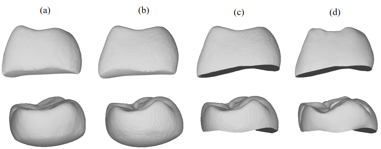
With the development of artificial intelligence, deep learning has been increasingly adopted for medical image analysis. [34, 37]. Several studies have leveraged deep learning algorithms to assist in dental crown design. Some of these approaches project IOS data from the occlusal perspective onto 2D depth maps, which are then used with image generation algorithms to create the intricate occlusal surface details of the crown [36, 43, 16]. However, these methods suffer from an intrinsic limitation: information loss introduced by the projection. Some crucial anatomical regions, such as the cervical margin, are obscured in 2D projections. The 2D depth projection is inherently incomplete, as it lacks data for regions obscured from the occlusal view. Post-processing is required to complete these areas. To address these issues, some recent studies have attempted to utilize point cloud completion networks combined with surface reconstruction algorithms for crown generation [12, 13, 42]. Nonetheless, such approaches increase the learning complexity, and there is an inherent trade-off between minimizing output noise and enhancing the details of the generation results. Moreover, the surface reconstruction algorithm in previous studies inherently produces watertight meshes [24, 27]. This process is fundamentally incompatible with a dental crown, which is an open genus-zero mesh. As illustrated in Figure 2, there is a noticeable overextension at the bottom of the reconstructed crown surface. These limitations constrain the clinical applicability of crown generation algorithms.
In this work, we proposed a Margin Aware Dental Crown generation framework (MADCrowner) for personalized dental crown design, primarily composed of two modules: CrownSegger and CrownDeformR. The CrownSegger combines both point cloud and volumetric information to segment the prepared abutment region from IOS data, thus accurately determining the cervical margin of the target abutment. In CrownDeformR, the output of CrownSegger and the IOS data are integrated as anatomical context features via a multiscale point cloud encoder. CrownDeformR utilizes these features to predict the deformation from a corresponding point cloud template to the target crown from coarse to fine. The surface of the target crown is generated from the deformed template using Differentiable Poisson Surface Reconstruction (DPSR). A postprocessing algorithm is applied to excise extraneous regions from the reconstructed surface, guided by the cervical margin line detected by CrownSegger.
Our main contributions are summarized as follows:
-
1.
We proposed MADCrowner, a novel cervical margin aware framework for dental crown design. The framework consists of two learning-based components, CrownSegger and CrownDeformR. MADCrowner improves the generated crowns significantly in both the accuracy and geometric details.
-
2.
We designed CrownSegger, a compact network for point cloud segmentation, to detect the cervical margin of the target abutment and impose the location of the cervical margin into CrownDeformR. The detected cervical margin is also utilized by a tailored post-process to eliminate the superfluous area generated by surface reconstruction, as shown in Figure 2.
-
3.
We introduced a concise network, CrownDeformR, for crown generation. CrownDeformR produces the target crown by deforming and refining the template mesh from coarse to fine. The output of CrownSegger is leveraged to enhance the performance.
-
4.
We established a large-scale intraoral scan dataset from clinical cases and conducted extensive experiments to evaluate the robustness and effectiveness of the proposed method.
2 Related Works
In this section, we introduce previous studies on the application of artificial intelligence in dental crown design, as well as research related to point cloud segmentation and mesh completion in general computer vision tasks, which can be applied to complete restoration design.
2.1 Deep Learning Methods for Dental Crown Design
CAD systems have become ubiquitous in restorative dental crown design, significantly boosting the efficiency of dental technicians. However, compared to other clinical applications, the level of automation and intelligence in current CAD systems for dental crown design remains relatively primitive. Driven by the rapid development of deep learning algorithms, several studies on dental crown generation have emerged in recent years [36, 51, 12, 13, 42].
[36] addressed dental crown generation by recasting it as a 2D image generation task. They projected IOS data as 2D depth maps from the occlusal perspective and generated the occlusal surfaces of the target crown with a conditional GAN (cGAN) [16, 8]. A series of post-processing steps was subsequently employed to reconstruct the final crown mesh from the generated occlusal surfaces. [51] demonstrated the feasibility of dental crown generation using point cloud completion networks by masking a single tooth from IOS data and reconstructing it via a point cloud completion model. [42] proposed a crown generation method based on a Point Transformer architecture. Their model takes IOS point clouds as input and generates the corresponding crown point cloud, which is then converted into a mesh using a “Shape as Points” (SAP) module. However, this method exhibits limited capability in reconstructing fine-grained anatomical details of dental crowns, such as grooves and fossae. [12] utilized AdaPointr [46], an advanced point completion network, to generate the dental crown and further modified the loss function to better suit the specific requirements of crown generation. This method demonstrated superior performance compared to similar existing approaches, achieving state-of-the-art results in dental crown generation.
2.2 Point Cloud Segmentation
According to the approaches used to process point cloud data, point cloud segmentation algorithms can be categorized into point-wise and voxel-wise methods. PointNet [30] and PointNet++ [31] are seminal works of the point-wise approach. However, their pooling-based feature aggregation limits the ability to capture the spatial correlation among points. PointTransformer [49] addressed this issue by employing self-attention blocks for adaptive feature learning. However, it requires a large-scale training dataset to achieve satisfactory generalization performance. For the voxel-wise methods, such as MinkowskiNet [5], the point cloud is voxelized as a 3D volume. Networks designed for 3D image segmentation are applied to process this discrete 3D volume. The point cloud segmentation is obtained by reversing the voxelization process to map the volume segmentation back to the point cloud. Voxel-wise methods primarily suffer from two main drawbacks: quantization loss caused by voxelization and substantial computational consumption required by 3D convolutions. Some voxel-wise methods utilize sparse convolutions [22] to reduce VRAM usage and improve computational efficiency. In recent years, several studies, such as PVCNN [23], have emerged that integrate point-wise and voxel-wise features for point cloud segmentation. These hybrid approaches combine the strengths of both methodologies, demonstrating excellent performance on point cloud segmentation tasks.
2.3 Point Cloud Completion and Surface Reconstruction
Due to their unique data structures, existing deep neural networks struggle to process mesh data directly. Consequently, the task of mesh completion is typically divided into two sub-tasks: point cloud completion and surface reconstruction. This decomposition enables the leveraging of deep neural networks for mesh completion. Previous research in point cloud completion has proposed many effective models, including PCN [47], FoldingNet [44], TopNet [35], and GRnet [41]. However, there is a critical limitation for these methods: the absence of normal vectors for their output points, which are an essential component for most classical mesh reconstruction algorithms. To bridge this gap between point cloud completion and mesh reconstruction, SAP [27] introduced a novel Differentiable Poisson Surface Reconstruction (DPSR) algorithm and integrated it with a point cloud completion network for the direct surface reconstruction from the point cloud. SAP has been proven to be a noise-robust mesh reconstruction approach, capable of reconstructing surface meshes from noisy point clouds. However, similar to traditional surface reconstruction algorithms, such as Poisson Surface Reconstruction (PSR) [19], SAP generates watertight meshes. This inherent characteristic directly contradicts the desired open, genus-zero morphology of dental crown meshes. This critical discrepancy has received only limited attention in prior research.


3 Methods
The overall pipeline of MADCrowner is illustrated in Figure 3. Given a target abutment, the cervical margin is first identified by CrownSegger to extract the cervical margin. Conditioned on this margin line and the IOS, CrownDeformR learns to model detailed dental anatomy via attention mechanism. By attending to the spatial relationships of adjacent and antagonist teeth, the network adaptively deforms an initial template crown which serves as a geometric prior to facilitate stable training. This data-driven deformation produces a crown point cloud that satisfies functional occlusion and proximal contact requirements. A watertight crown mesh is subsequently reconstructed using SAP. Due to the watertight constraint imposed by SAP, topological artifacts (e.g., spurious mesh extensions near the cervical boundary) may arise. A dedicated post-processing module is therefore applied to remove these reconstruction-induced artifacts, ensuring compliance with clinical requirements while preserving the learned anatomical morphology.
3.1 CrownSegger
The CrownSegger, illustrated in Figure 4, is a hybrid point-voxel framework for point cloud segmentation. The initial point-wise features are constructed by concatenating the spatial coordinates and normal vectors. The input point cloud is voxelized into a volume, where each voxel may contain multiple points. Voxel-wise features are then generated by aggregating point-wise features within each voxel through cascaded voxel feature encoding (VFE) layers [50]. These sparse voxel-wise features are transformed into a dense 3D volume, as shown in Figure 4 (b), and processed by a VNet [1] backbone to capture multi-scale anatomical patterns. We concatenate the voxel-wise features from the output feature volume with their corresponding point-wise features, as illustrated in Figure 4 (c), and fuse them with an MLP to predict the segmentation results. The training of CrownSegger is supervised by cross-entropy loss and dice loss. Finally, the cervical margin of the target abutment is identified by extracting and smoothing the boundary of the prepared abutment.
3.2 CrownDeformR
The overall architecture of our CrownDeformR is illustrated in Figure 5. The IOS and the segmentation of the prepared abutment are first encoded to extract geometric and contextual features. Following a CAD-inspired crown design paradigm, CrownDeformR generates the target crown by deforming the corresponding tooth template conditioned on the extracted features. The template is progressively refined in a coarse-to-fine manner to produce the final crown geometry.
3.2.1 Network Primitives
The proposed architecture consists of three specialized Transformer-based modules: the Geometry Aware Transformer (GAT), the Self Attention Transformer (SAT), and the Cross Attention Transformer (CAT). The GAT performs hierarchical feature extraction with progressive downsampling while preserving local geometric structures, enabling robust representation of fine-grained dental details. Within each resolution scale, the SAT is employed to model long-range dependencies within feature tokens, enhancing internal contextual interactions and enabling globally consistent representations across different stages of the framework. To guide crown generation conditioned on the IOS context, the CAT models the spatial correspondence between crown queries and IOS features, capturing their relative positional dependencies and geometric interactions. The detailed mathematical formulations and tensor operations of these modules are provided in Figure 18 and Figure 19 of the Appendix.
3.2.2 Context-Aware Feature Extraction
In the initial feature encoding stage, the IOS point cloud is augmented with the abutment segmentation label to provide explicit margin guidance. This hybrid input is processed through a sequence of GAT-SAT blocks, which yields a global context vector along with multi-scale intermediate features . This hierarchical encoding scheme ensures that the subsequent generative stages are informed by both the global dental alignment and localized margin details.
3.2.3 Hierarchical Template Deformation and Refinement
Inspired by clinical CAD workflows, CrownDeformR deforms an initial template selected via the FDI notation system through a two-stage process. During the initial deformation stage, a CAT block captures the spatial dependencies between the template and the global IOS features, guiding the coarse template deformation. The resulting features are further processed by SAT blocks to predict the coarse crown point cloud . Subsequently, in the multi-scale refinement stage, this coarse result is progressively upsampled through two refinement modules. Each module leverages CAT to cross-reference localized IOS features, enabling the network to actively learn and reconstruct fine-grained anatomical details, such as occlusal grooves and proximal contact points.
3.3 Optimization Objectives
The CrownDeformR is optimized using a hierarchical loss function that enforces geometric fidelity from a coarse-to-fine perspective. To ensure the generated crown captures both stable dental proportions and fine-grained anatomical details, we implement a deep supervision strategy across multiple resolutions. Specifically, for the initial deformation and the first refinement stages, we utilize standard Chamfer Distance (CD) to supervise the point cloud at different scales. The coarse loss () is calculated between the coarse point cloud and a downsampled ground truth to establish the basic spatial positioning and crown proportions. Subsequently, the first refinement loss () supervises the downsampled ground truth , ensuring structural consistency as the point density increases. While these global objectives provide a stable optimization trajectory, we utilize the Curvature and Margin Penalty Loss (CMPL, ) for the final refinement stage to capture high-frequency anatomical features such as occlusal fossae and ensure precise marginal fit. Unlike vanilla CD, CMPL explicitly prioritizes high-curvature regions and the cervical boundary, formulated as:
| (1) | |||
where denotes the final predicted point cloud, represents the normalized curvature, and refers to the set of points on the ground-truth margin line. The exponential term adaptively increases the penalty in areas with complex anatomy, while the indicator function enforces strict alignment with the prepared abutment. Finally, the surface reconstruction is supervised by an MSE loss () on the DPSR grid. The total loss is the sum of all aforementioned components. We discuss detailed technical specifications, including the loss function formulations of , , and specific tensor shape transformations at each stage in the Appendix.
3.4 Post-processing for Reconstructed Mesh
Most of the surface reconstruction algorithms, including DPSR, produce meshes that are inherently watertight. However, the dental crown is an open genus-zero mesh. It is necessary to eliminate the overextension from the reconstructed mesh to obtain the final result, a step that is rarely mentioned in previous research [12, 13, 42]. We design a tailored post-processing procedure to address this issue. The cervical margin is identified as the boundary of the prepared abutment, which is predicted by CrownSegger. The points on the margin line are smoothed with B-spline kernels. These smoothed points, along with their centroid, define a surface whose normal vectors align with the tooth growth direction. Faces within the reconstructed mesh that are below this surface are then removed. To ensure the mesh boundary precisely conforms to the margin line, points on the boundary of the processed mesh are projected onto the smoothed margin line. Following these post-processing steps, the reconstructed crown surface is transformed into an open genus-zero mesh, which can be manipulated within CAD systems for clinical application.
4 Experiments and Results
Input:
Mesh:
Point-wise labels for the target abutment:
Output:
Cervical margin vertex set:
Step 1: Abutment Extraction
Extract faces from , in which three vertices are labeled as abutment:
.
Step 2: Noise Exclusion
Construct a sub-mesh .
Extract the largest connected component from .
Step 3: Boundary Detection
Extract the vertices in the boundary of .
Step 4: Cervical Margin Estimation
Estimate the cervical margin by B-spline interpolation with .
Resample 1,000 vertices () uniformly on the cervical margin.
return
4.1 Datasets
4.1.1 Dataset for prepared abutment segmentation
We built a dataset for prepared abutment segmentation that consists of 576 IOS data. Each IOS is point-wise annotated, indicating whether individual points belong to the prepared abutment. Among these, 476 scans are allocated for training, and 100 for testing. The cervical margin, which is defined as the boundary of the prepared abutment, is extracted by Algorithm 1.
4.1.2 Dataset for Dental Crown Design
| Jaw Side | Tooth Position | Number of Cases |
| Upper | 1st Premolar | 543 |
| 2nd Premolar | 412 | |
| 1st Molar | 1096 | |
| 2nd Molar | 270 | |
| Lower | 1st Premolar | 152 |
| 2nd Premolar | 178 | |
| 1st Molar | 1452 | |
| 2nd Molar | 499 |
We collect raw IOS data acquired from 4,602 patients with single tooth defects, in which the target teeth are prepared as abutments. For each patient, the corresponding dental crown designed by dental technicians is also collected. The target teeth in the dataset encompass all the premolars and molars. For each IOS data sample, we extract a 2 cm-sided cubic region centered at the centroid of its corresponding crown. The dataset is divided into training, validation, and testing sets with 8:1:1. We utilize stratified sampling among the types of target teeth, ensuring that the distribution of target teeth is consistent across both the training and test sets. To enhance computational efficiency during training, we pre-extract the curvature for each vertex in the IOS data, as well as the boundaries of the IOS data, specifically within the test set.
4.2 Implementation Details
The CrownSegger was implemented with PyTorch and trained on a NVIDIA GTX 3080 Ti with a batch size of 16 for 300 epochs. We used the AdamW optimizer with an initial learning rate of to update the network parameters and applied a cosine learning rate scheduler during training.
The CrownDeformR was also implemented with PyTorch and trained for 500 epochs on 4 NVIDIA RTX 4090 GPUs with a batch size of 8. Configurations of the optimizer and learning rate scheduler for CrownDeformR are the same as CrownSegger. The abutment mask used by CrownDeformR is the zero-shot prediction from CrownSegger. The employed transformer blocks (GAT, SAT, and CAT) leveraged multi-head attention with 4 heads and 512 hidden channels. The training of CrownDeformR is supervised by the combination of the aforementioned loss functions:
| (2) |
The dental crown generation pipeline consists of two distinct models: CrownSegger and CrownDeformR. During the inference stage, these two models are combined as a unified network, MADCrowner, which comprises 37.3 M parameters. MADCrowner requires only 1.1 GB VRAM for the inference of a single sample. The single-inference latency is approximately 600 ms (Segmentation, Generation and Post-processing) on an NVIDIA L20 GPU. This resource frugality makes the MADCrowner an ideal candidate for on-site deployment.
| Methods | CD-L2 () | Fidelity Distance () | Hausdorff Distance (mm) | F-score | ||||||||
| Premolar | Molar | Overall | Premolar | Molar | Overall | Premolar | Molar | Overall | Premolar | Molar | Overall | |
| PCN[47] +SAP | 0.271 | 0.318 | 0.305 | 0.118 | 0.148 | 0.140 | 1.400 | 1.458 | 1.441 | 0.850 | 0.889 | 0.878 |
| TopNet[35] +SAP | 0.354 | 0.409 | 0.394 | 0.164 | 0.200 | 0.190 | 1.752 | 1.869 | 1.836 | 0.770 | 0.812 | 0.800 |
| GRnet[41] +SAP | 0.230 | 0.254 | \cellcolorsecond0.247 | 0.110 | \cellcolorsecond0.132 | 0.126 | \cellcolorsecond1.118 | \cellcolorsecond1.139 | \cellcolorsecond1.134 | \cellcolorsecond0.874 | \cellcolorsecond0.901 | \cellcolorsecond0.894 |
| DMCv2 [12] | \cellcolorsecond0.227 | 0.264 | 0.254 | \cellcolorsecond0.107 | \cellcolorsecond0.132 | \cellcolorsecond0.125 | 1.227 | 1.252 | 1.245 | 0.868 | 0.897 | 0.888 |
| MADCrowner | \cellcolorbest0.176 | \cellcolorbest0.174 | \cellcolorbest0.175 | \cellcolorbest0.098 | \cellcolorbest0.092 | \cellcolorbest0.094 | \cellcolorbest1.093 | \cellcolorbest0.996 | \cellcolorbest1.027 | \cellcolorbest0.903 | \cellcolorbest0.932 | \cellcolorbest0.924 |
| Methods | CD-L2 () | Fidelity Distance () | Hausdorff Distance (mm) | F-score | ||||||||
| Premolar | Molar | Overall | Premolar | Molar | Overall | Premolar | Molar | Overall | Premolar | Molar | Overall | |
| GRnet+SAP [41] | 0.212 | 0.210 | 0.210 | 0.111 | 0.100 | 0.103 | 1.212 | 1.307 | 1.280 | 0.895 | 0.901 | 0.899 |
| DMCv2 [12] | 0.259 | 0.251 | 0.253 | 0.137 | 0.123 | 0.127 | 1.329 | 1.319 | 1.322 | 0.868 | 0.875 | 0.873 |
| VBCD [40] | 0.212 | 0.207 | 0.209 | 0.108 | 0.109 | 0.109 | 1.115 | 1.163 | 1.150 | 0.896 | 0.915 | 0.909 |
| Diffusion SDF [4] | 0.228 | 0.215 | 0.219 | 0.116 | 0.108 | 0.110 | 1.216 | 1.458 | 1.390 | 0.875 | 0.900 | 0.893 |
| MADCrowner | \cellcolorbest0.198 | \cellcolorbest0.180 | \cellcolorbest0.185 | \cellcolorbest0.083 | \cellcolorbest0.087 | \cellcolorbest0.086 | \cellcolorbest1.096 | \cellcolorbest1.026 | \cellcolorbest1.046 | \cellcolorbest0.907 | \cellcolorbest0.920 | \cellcolorbest0.917 |
| MADCrowner w/o Postprocessing | 0.556 | 1.186 | 1.006 | 0.086 | 0.094 | 0.091 | 3.176 | 4.342 | 4.016 | 0.826 | 0.831 | 0.830 |

4.3 Abutment mask Segmentation
In this section, we conducted a comparative analysis of CrownSegger with several widely adopted point cloud segmentation algorithms, evaluating their performance on both target abutment segmentation and cervical margin extraction. The cervical margin of the target abutment is extracted from its segmentation mask.
For the abutment segmentation, we employed accuracy and Intersection over Union (IoU) as quantitative metrics. The performance of cervical margin extraction was evaluated using the Hausdorff Distance (HDF distance), calculated in the physical coordinate system.
We compared CrownSegger with several prevalent point cloud segmentation models, including PointNet [30], PointNet++ [31], and PointTransformer [49], on our dataset for abutment segmentation. CrownSegger consistently outperformed these methods across all the evaluated metrics. The improvement in HDF distance is particularly significant, decreasing from 1.237 mm to 0.328 mm (Table 2). The performance of cervical margin extraction was also evaluated on the dataset for dental crown design. For this dataset, the ground truth of the cervical margin was defined as the boundary of the dental crown. The cervical margin derived from the zero-shot prediction of CrownSegger achieved remarkable performance with an average HDF distance of 0.575 mm, indicating excellent robustness and generalization ability of CrownSegger.
Figure 6 visualizes an example of the prepared abutment segmentation and the extracted cervical margin. As shown in this figure, CrownSegger demonstrated better segmentation performance, with notably fewer misclassified points. Additionally, the cervical margin extracted from CrownSegger’s prediction also exhibited better consistency with the ground truth.
It should be noted that CrownSegger is constructed with a transformer-free backbone, which grants it distinct advantages in computational efficiency and memory usage. This makes the model particularly suitable for clinical deployment on edge computing devices, as it achieves high performance with minimal hardware resource demands.
4.4 Dental Crown Generation
4.4.1 Evaluation Metric
To validate the effectiveness of MADCrowner, we conducted extensive comparative experiments against existing dental crown generation methods and ablation studies. We used the L2-normed Chamfer Distance (CD-L2), fidelity distance [41], F-score [13], and HDF distance for quantitative assessment of experimental results from different perspectives. CD-L2, fidelity distance, and F-score measure the overall similarity between the generated crown and the ground truth. In contrast, the HDF distance quantifies the maximum deviation of the generated crown. HDF distance is a critical evaluation metric for the generated crowns, as it provides an intuitive assessment of their compatibility with the corresponding IOS data. In this section, all distance-related metrics were calculated in the physical coordinate system measured in millimeters (mm).
4.4.2 Experimental Result


We compared the performance of MADCrowner with several advanced point cloud completion models, including PCN [47], TopNet [35], and GRnet [41], which were integrated with SAP [27] to reconstruct the corresponding mesh. Additionally, DMCv2 [12], the state-of-the-art dental crown generation method, was also evaluated as a comparative experiment. Following the previous works [13, 12], quantitative metrics were evaluated on the generated point clouds. The experimental results are listed in Table 3. MADCrowner achieved the best performance across all the metrics. Specifically, our method surpassed the second-highest performance in CD-L2, fidelity distance, and F-score by 29.2%, 26.4%, and 3.36%, respectively. This demonstrates that MADCrowner significantly enhanced the consistency between the generated dental crowns and the ground truth. In terms of HDF distance, our method achieved a significant improvement by reducing the best performance from 1.139 mm to 1.027 mm. This notable reduction in HDF distance indicates that our approach generated dental crowns with less noise and lower deviation from the ground truth. We further conducted a quantitative evaluation for the reconstructed dental crown mesh and incorporated several voxel-based methods [40, 4] for comparison. The evaluation results are shown in Table 4, where MADCrowner consistently achieved the best performance across all metrics. Compared to Table 3, there are slight discrepancies in the quantitative metrics of a single method when measured on the generated point cloud versus the reconstructed mesh. These gaps are induced by the inherent perturbations of the mesh reconstruction algorithm and the effect of the postprocessing.
We exhibited visual comparison of MADCrowner to point cloud-based methods and voxel-based methods in Figure 7 and Figure 8, respectively. The surface distance error of these experimental results is visualized using a colormap. It is obvious that MADCrowner yields the highest consistency between the generated crowns and the ground truth. Additionally, it can be observed that the crowns generated by our method exhibit remarkable improvement in intricate details, such as grooves and fossae.
4.5 Ablation Study and Cost-Benefit Discussion
| Components | CD-L2 () | Fidelity Distance () | Hausdorff Distance (mm) | F-Score | Efficiency | |||||||||||
| Cervical Margin Constraints | Initial Crown Template | Crown Template Deformation | Premolar | Molar | Overall | Premolar | Molar | Overall | Premolar | Molar | Overall | Premolar | Molar | Overall | VRAM (MB) | Inference Time (ms) |
| ✗ | ✗ | ✗ | 0.214 | 0.222 | 0.220 | 0.106 | 0.114 | 0.112 | 1.419 | 1.347 | 1.367 | 0.866 | 0.905 | 0.895 | \cellcolorbest1229 | \cellcolorbest87 |
| ✗ | ✓ | ✗ | 0.204 | 0.217 | 0.213 | 0.102 | 0.110 | 0.107 | 1.372 | 1.291 | 1.314 | 0.878 | 0.910 | 0.901 | 1229 | 90 |
| ✗ | ✗ | ✓ | 0.215 | 0.211 | 0.212 | 0.115 | 0.106 | 0.108 | 1.365 | 1.280 | 1.301 | 0.882 | 0.911 | 0.903 | 1305 | 100 |
| ✗ | ✓ | ✓ | 0.188 | 0.195 | 0.193 | 0.099 | 0.102 | 0.101 | 1.182 | 1.152 | 1.160 | 0.886 | 0.921 | 0.911 | 1305 | 103 |
| ✓ | ✓ | ✓ | \cellcolorbest0.176 | \cellcolorbest0.174 | \cellcolorbest0.175 | \cellcolorbest0.098 | \cellcolorbest0.092 | \cellcolorbest0.094 | \cellcolorbest1.093 | \cellcolorbest0.996 | \cellcolorbest1.027 | \cellcolorbest0.903 | \cellcolorbest0.932 | \cellcolorbest0.924 | 1305 | 105 |
The efficacy of CrownDeformR hinges upon three critical components: the initial crown template, the template deformation module, and the cervical margin constraints. In this section, we demonstrate the contribution of these components with a series of ablation studies. The results of the ablation studies are listed in Table 5. We visualized the results of the ablation studies with an example in Figure 9. The dental crown generated by the full CrownDeformR demonstrated superior performance in both overall similarity and intricate details compared to the control groups. Beyond the quantitative performance improvements, we further perform a cost-benefit analysis to evaluate the practical necessity of each component. Although the numerical gains introduced by the additional modules may appear modest, such improvements are clinically meaningful in dental crown restoration. Given the high precision requirements of occlusal fitting, even sub-millimeter reductions in geometric deviation can substantially improve patient comfort and reduce the need for chairside adjustments. Meanwhile, the added computational overhead remains marginal. The incorporation of the template deformation module and cervical margin constraints increases GPU memory consumption (VRAM) by less than 100 MB and inference time by less than 30 milliseconds. Considering this negligible efficiency cost relative to the clinically significant accuracy gains, the complete CrownDeformR framework is both practically justified and necessary for high-fidelity dental crown generation.
4.5.1 Initial crown template
Inspired by the CAD workflow of dental crown design by technicians, CrownDeformR selects an appropriate initial template based on the FDI label of the target tooth. This template is then deformed and refined by CrownDeformR to generate the final crown. To evaluate the impact of the specified initial crown template, we conducted an ablation study in which the initial crown template was replaced with a hemisphere of 7.5 mm radius. The results presented in the first and second rows of Table 5 demonstrate that an appropriate initial crown template can improve the generation results across all the metrics.
4.5.2 Crown Template Deformation
In CrownDeformR, the initial crown template is processed by a template deformation module to create a coarse crown. This coarse representation is then refined to produce the final dental crown. As demonstrated by a comparison of the first and third rows in Table 5, the integration of the template deformation module significantly enhances overall crown generation performance compared to the direct utilization of the initial template as an input for the refinement module.
4.5.3 Cervical Margin Constraints
The cervical margin serves as a critical reference in dental crown design. In clinical practice, dental technicians begin the crown design by annotating the cervical margin on the prepared abutment. This delineation then guides the subsequent design process, ultimately defining the boundary of the dental crown. CrownDeformR also incorporates cervical margin constraints. To enhance awareness of the cervical margin, CrownDeformR leverages the prediction of CrownSegger as part of the input, in addition to the IOS data. A comparison of the last two rows in Table 5 reveals that incorporating the cervical margin constraint significantly enhances all evaluated metrics. Notably, the improvement in HDF distance is particularly pronounced. This is consistent with the fact that the maximum deviation in generated crowns often occurs in the margin region. It suggests that the cervical margin constraint enhances the quality of the generated crown in the critical cervical region. As shown in Figure 10, the application of cervical margin constraints improves the consistency between the generated point cloud and the ground truth. This enhancement is especially notable near the cervical margin.
4.5.4 Hyper-parameters for CMPL
As described in the section CrownDeformR, CrownDeformR was trained with CMPL to enhance the fidelity of crown details and the peri-cervical regions. CMPL is composed of two distinct parts: Curvature Penalty Loss (CPL) and Margin Penalty Loss (MPL):
is the scale factor of curvature weights. We conducted an ablation study to find the optimal for dental crown generation. As shown in Figure 11, both CD-L2 and Hausdorff Distance achieved the best performance when is set to 1. It was adopted as the experimental setting in this work.


4.5.5 Mesh Reconstruct Methods
In this section, we evaluate the effects of different mesh reconstruction algorithms. For comparative analysis, we utilized Neural Kernel Surface Reconstruction (NKSR) [14] and Point2Mesh [9] to reconstruct surfaces from the point clouds generated by MADCrowner. The comparison results are exhibited both visually and quantitatively, in Figure 13 and Table 6. It is evident that the performance of our reconstruction method surpassed other approaches.
| Methods | CD-L2 () | Fidelity Distance () | Hausdorff Distance (mm) | F-score | ||||||||
| Premolar | Molar | Overall | Premolar | Molar | Overall | Premolar | Molar | Overall | Premolar | Molar | Overall | |
| MADCrowner (Point2Mesh) | 0.247 | 0.238 | 0.240 | 0.129 | 0.145 | 0.140 | 1.841 | 1.832 | 1.834 | 0.842 | 0.849 | 0.847 |
| MADCrowner (NKSR) | 0.235 | 0.227 | 0.229 | 0.132 | 0.126 | 0.128 | 1.568 | 1.485 | 1.508 | 0.857 | 0.863 | 0.861 |
| Ours | \cellcolorbest0.198 | \cellcolorbest0.180 | \cellcolorbest0.185 | \cellcolorbest0.083 | \cellcolorbest0.087 | \cellcolorbest0.086 | \cellcolorbest1.096 | \cellcolorbest1.026 | \cellcolorbest1.046 | \cellcolorbest0.907 | \cellcolorbest0.920 | \cellcolorbest0.917 |

4.6 Proximal Contact Analysis
Appropriate proximal contacts with adjacent teeth are a critical consideration in dental crown design. Excessive contact can lead to periodontal ligament damage, while overly loose contacts may result in food impaction. To address these concerns, dental technicians commonly incorporate appropriate intersection regions between the crown and adjacent teeth. This intentional design provides clinicians with the necessary flexibility to make precise adjustments during crown restoration, ensuring optimal proximal contacts of the target crown. To quantitatively assess the fidelity of proximal contacts, we calculated the proximal intersection area (PIA) between the dental crown and its adjacent teeth. The deviation in PIA between the generated crown and the ground truth then served as the metric for evaluating the accuracy of the proximal contacts for the generated results. Table 7 shows that the results of MADCrowner exhibit the lowest error, which indicates that the dental crowns produced by MADCrowner maintain excellent consistency with the ground truth in proximal contacts. As depicted in Figure 12, the proximal contact regions of the generated crowns exhibit excellent fidelity with the ground truth in both premolars and molars. Compared with VBCD, the model with the strong geometric performance in Table 4, our method produces proximal contact distributions that are more consistent with the ground truth in terms of spatial localization and contact extent.
4.7 Failure Case Analysis
The MADCrowner demonstrated strong generalization ability and produced high-quality crown meshes for the majority of cases in the test set. However, beyond isolated extreme outliers, we observed several recurring failure patterns associated with specific clinical conditions. Figure 14 presents representative failure cases generated by MADCrowner. Our analysis reveals that crown generation is more prone to failure under the following conditions: (1) inadequate tooth preparation, where irregular or insufficient reduction compromises geometric inference; (2) incomplete intraoral scans, particularly holes or missing regions in adjacent teeth; and (3) absence of adjacent or antagonist teeth, which limits accurate reconstruction of occlusal relationships. In these scenarios, the generated crowns may exhibit improper occlusal contacts or suboptimal morphological adaptation. The identified patterns are consistent with clinical experience, highlighting the importance of standardized tooth preparation and complete adjacent and antagonist tooth information for reliable automated crown design.


5 Discussion and Conclusion
In this work, we propose a novel dental crown generation framework, MADCrowner, which primarily consists of CrownSegger and CrownDeformR. The IOS data is first processed using CrownSegger to generate a segmentation mask of the prepared abutment, from which the cervical margin is subsequently extracted. CrownDeformR integrates the IOS data and the abutment mask to generate the dental crown for the target tooth. Our approach achieved superior performance across all the evaluated metrics, including CD-L2, Fidelity Distance, HDF Distance, and F-score. Compared to the approximately 15 minutes required for a dental technician to manually design a crown using a CAD system, MADCrowner generates a dental crown within 500 ms. This dramatic acceleration significantly enhances the efficiency of dental technicians.
For dental crown generation methods based on point cloud completion networks, a uniform spatial distribution and sufficient density of the output point cloud are crucial for accurate dental crown generation. Figure 15 compares the point clouds generated by DMC v2, GRnet, and MADCrowner. Both DMC v2 and GRnet fail to generate the anatomical details of the occlusal surface, such as grooves and fossae. GRnet voxelizes the point cloud, which inherently limits the output point cloud density due to the spatial resolution constraints of the input volume. DMC v2 addresses this insufficient point cloud density by directly generating point proxies from IOS data using a point transformer encoder. However, the encoder of DMC v2 and similar methods [38, 45, 46] is built upon self-attention blocks. All the features are derived exclusively from the IOS data. This often leads to an inhomogeneous spatial distribution of the generated point cloud. To solve this limitation, CrownDeformR employed cross-attention blocks to generate point proxies, which integrate features from the IOS data with the spatial information from the specific initial crown template. As shown in Figure 15, CrownDeformR generates point clouds with a uniform spatial distribution and excellent occlusal surface details.
It should be noted that meshes produced by most of surface reconstruction algorithms are inherently watertight. This property introduces overextension in the reconstructed crown, a challenge that has rarely been discussed in previous studies. As illustrated in Figure 16, MADCrowner addresses this issue by incorporating a post-processing method that trims these extraneous areas with reference to the cervical margin. In addition, the post-processing algorithm also guarantees accurate consistency between the margins of the generated crown and the ground truth.
Besides proximal contacts, appropriate occlusal contacts are also crucial for dental crown design. MADCrowner produces dental crowns with favorable occlusal contacts. As depicted in Figure 17, which visualizes the occlusal contacts for two typical crowns, the distribution of occlusal contact regions on the generated crowns reveals great similarity with the ground truth. Compared with VBCD, a method that achieves the best performance in Table 4, our method produces occlusal contact regions whose spatial distribution and contact intensity patterns more closely resemble those of the ground truth.
While our experimental results demonstrate that MADCrowner outperforms existing methods, certain limitations persist. Dental crown generation is an inherently open-ended task. Dental technicians often design noticeably different crowns for the same target tooth, reflecting the diverse range of clinically acceptable solutions. However, current methodologies, including ours, typically constrain the predicted crown shape through regression-based losses, such as the Chamfer Distance. This approach inadvertently encourages the network to produce a “smoothed average” of plausible outputs rather than capturing the true “mode” of the underlying distribution. Consequently, the generated crowns may lack critical anatomical details, such as grooves and fossae, which are essential for optimal function and aesthetics.
Future work should explore the incorporation of advanced generative models, such as diffusion models [10], within 3D crown generation to produce high-quality crowns with richer morphological details. Moreover, expansion of the IOS dataset is also crucial to further enhance and validate the generalization capability of crown generation algorithms. This expansion should particularly focus on cases in which the incisor or canine is the target tooth.
CRediT authorship contribution statement
Linda Wei:Methodology, Validation, Visualization, Writing original draft. Chang Liu:Methodology, Writing original draft. Wenran Zhang:Writing original draft, Visualization. Yuxuan Hu:Writing original draft. Ruiyang Li:Writing original draft. Feng Qi: Writing original draft. Changyao Tian:Writing original draft. Ke Wang:Writing original draft. Yuanyuan Wang:Project administration, Supervision, Writing review & editing. Shaoting Zhang:Project administration, Supervision, Writing review & editing. Dimitris Metaxas:Project administration, Supervision Hongsheng Li: Project administration, Supervision, Writing review & editing, Funding Acquisition
Data availability
Due to privacy issues, the data used in this study is confidential.
Declaration of generative AI and AI-assisted technologies in the writing process.
During the preparation of this work, the authors used ChatGPT in order to correct grammatical errors. After using this tool, the authors reviewed and edited the content as needed and take full responsibility for the content of the published article.
Declaration of competing interest
The authors declare that they have no known competing financial interests or personal relationships that could have appeared to influence the work reported in this paper.
Acknowledgement
The authors would like to thank The Chinese University of Hong Kong, Fudan University, SenseTime, and Rutgers University for providing computational resources and methodology guidance. They also wish to express their appreciation to the Department of Second Dental Center at Shanghai Ninth People’s Hospital, Shanghai Jiao Tong University School of Medicine, and Shanghai Stomatological Hospital for their professional insights and support throughout the project. Finally, the authors are grateful to the Centre for Perceptual and Interactive Intelligence (CPII) under InnoHK and the Natural Science Foundation of Sichuan Province for their funding support.
Funding
This project was funded in part by the Centre for Perceptual and Interactive Intelligence (CPII) Ltd under the Innovation and Technology Commission (ITC)’s InnoHK initiative, and in part by the Guangdong Basic and Applied Basic Research Foundation (Grant No. 2023B1515130008, XW). This work was also supported in part by the National Natural Science Foundation of China (Grant No. 62271115), and in part by the Natural Science Foundation of Sichuan Province (Grant No. 2025ZNSFSC0455).
References
- [1] (2020) VNet: an end-to-end fully convolutional neural network for road extraction from high-resolution remote sensing data. Ieee Access 8, pp. 179424–179436. Cited by: §3.1.
- [2] (2023) Efficacy of cad/cam technology in dental procedures performed by students: a systematic scoping review of randomized clinical trials. Heliyon 9 (4). Cited by: §1.
- [3] (2025) Time efficiency and cost analysis between digital and conventional workflows for the fabrication of fixed dental prostheses: a systematic review. The Journal of Prosthetic Dentistry 133 (1), pp. 71–84. Cited by: §1.
- [4] (2023) Diffusion-sdf: conditional generative modeling of signed distance functions. In Proceedings of the IEEE/CVF international conference on computer vision, pp. 2262–2272. Cited by: §4.4.2, Table 4, Table 7.
- [5] (2019) 4d spatio-temporal convnets: minkowski convolutional neural networks. In Proceedings of the IEEE/CVF conference on computer vision and pattern recognition, pp. 3075–3084. Cited by: §2.2.
- [6] (2011) The use of cad/cam in dentistry. Dental Clinics of North America 55 (3), pp. 559–ix. Cited by: §1.
- [7] (2001) Tooth preparations for complete crowns: an art form based on scientific principles. The Journal of prosthetic dentistry 85 (4), pp. 363–376. Cited by: §1.
- [8] (2020) Generative adversarial networks. Communications of the ACM 63 (11), pp. 139–144. Cited by: §2.1.
- [9] (2020) Point2mesh: a self-prior for deformable meshes. arXiv preprint arXiv:2005.11084. Cited by: §4.5.5.
- [10] (2020) Denoising diffusion probabilistic models. Advances in neural information processing systems 33, pp. 6840–6851. Cited by: §5.
- [11] (2024) Basic guide to dental procedures. John Wiley & Sons. Cited by: §1.
- [12] (2025) Personalized dental crown design: a point-to-mesh completion network. Medical Image Analysis 101, pp. 103439. Cited by: §1, §2.1, §2.1, §3.4, §4.4.2, Table 3, Table 4, Table 7, B. Generalization on Incisors and Canines.
- [13] (2023) From mesh completion to ai-designed crown. In International Conference on Medical Image Computing and Computer-Assisted Intervention, pp. 555–565. Cited by: §1, §2.1, §3.4, §4.4.1, §4.4.2, B. Generalization on Incisors and Canines.
- [14] (2023) Neural kernel surface reconstruction. In Proceedings of the IEEE/CVF Conference on Computer Vision and Pattern Recognition, pp. 4369–4379. Cited by: §4.5.5.
- [15] (1984) Dentistry—designation system for teeth and areas of the oral cavity. ISO. Cited by: A.b Detail of CrownDeformeR.
- [16] (2017) Image-to-image translation with conditional adversarial networks. In Proceedings of the IEEE conference on computer vision and pattern recognition, pp. 1125–1134. Cited by: §1, §2.1.
- [17] (2016) CAD-cam the future of digital dentistry: a review. IP Ann Prosthodont Restor Dent 2 (2), pp. 33–6. Cited by: §1.
- [18] (2016) Time-efficiency analysis of the treatment with monolithic implant crowns in a digital workflow: a randomized controlled trial. Clinical oral implants research 27 (11), pp. 1401–1406. Cited by: §1.
- [19] (2013) Screened poisson surface reconstruction. ACM Transactions on Graphics (ToG) 32 (3), pp. 1–13. Cited by: §2.3.
- [20] (2021) Endodontic indications among patients visiting a tertiary care center: a descriptive cross-sectional study. JNMA: Journal of the Nepal Medical Association 59 (240), pp. 741. Cited by: B. Generalization on Incisors and Canines.
- [21] (2025) Impact of traumatic dental injuries on oral health-related quality of life among primary school children in dar es salaam. Clinical and Experimental Dental Research 11 (1), pp. e70105. Cited by: §1.
- [22] (2015) Sparse convolutional neural networks. In Proceedings of the IEEE conference on computer vision and pattern recognition, pp. 806–814. Cited by: §2.2.
- [23] (2019) Point-voxel cnn for efficient 3d deep learning. Advances in neural information processing systems 32. Cited by: §2.2.
- [24] (1998) Marching cubes: a high resolution 3d surface construction algorithm. In Seminal graphics: pioneering efforts that shaped the field, pp. 347–353. Cited by: §1.
- [25] (2009) A review of dental cad/cam: current status and future perspectives from 20 years of experience. Dental materials journal 28 (1), pp. 44–56. Cited by: §1.
- [26] (2022) Global oral health status report: towards universal health coverage for oral health by 2030. World Health Organization. Cited by: §1.
- [27] (2021) Shape as points: a differentiable poisson solver. Advances in Neural Information Processing Systems 34, pp. 13032–13044. Cited by: §1, §2.3, §4.4.2.
- [28] (2019) Oral diseases: a global public health challenge. The Lancet 394 (10194), pp. 249–260. Cited by: §1.
- [29] (2015) Tooth preparation for full-coverage restorations—a literature review. Clinical oral investigations 19, pp. 959–968. Cited by: §1.
- [30] (2017) Pointnet: deep learning on point sets for 3d classification and segmentation. In Proceedings of the IEEE conference on computer vision and pattern recognition, pp. 652–660. Cited by: §2.2, §4.3, Table 2.
- [31] (2017) Pointnet++: deep hierarchical feature learning on point sets in a metric space. Advances in neural information processing systems 30. Cited by: §2.2, §4.3, Table 2.
- [32] (2023) Clinical applications and mechanical properties of cad-cam materials in restorative and prosthetic dentistry: a systematic review. Journal of functional biomaterials 14 (8), pp. 431. Cited by: §1.
- [33] (2011) Frequency and distribution of teeth requiring endodontic therapy in an argentine population attending a specialty clinic in endodontics. International dental journal 61 (5), pp. 257–260. Cited by: B. Generalization on Incisors and Canines.
- [34] (2017) Deep learning in medical image analysis. Annual review of biomedical engineering 19 (1), pp. 221–248. Cited by: §1.
- [35] (2019) Topnet: structural point cloud decoder. In Proceedings of the IEEE/CVF conference on computer vision and pattern recognition, pp. 383–392. Cited by: §2.3, §4.4.2, Table 3, Table 7.
- [36] (2021) DCPR-gan: dental crown prosthesis restoration using two-stage generative adversarial networks. IEEE Journal of Biomedical and Health Informatics 26 (1), pp. 151–160. Cited by: §1, §2.1, §2.1.
- [37] (2024) SenseCare: a research platform for medical image informatics and interactive 3d visualization. Frontiers in Radiology 4, pp. 1460889. Cited by: §1.
- [38] (2024) Pointattn: you only need attention for point cloud completion. In Proceedings of the AAAI Conference on artificial intelligence, Vol. 38, pp. 5472–5480. Cited by: §5, A.b Detail of CrownDeformeR.
- [39] (1994) Relative frequency of teeth needing endodontic treatment in 3350 consecutive endodontic patients. Journal of Endodontics 20 (8), pp. 399–401. Cited by: B. Generalization on Incisors and Canines.
- [40] (2025) VBCD: a voxel-based framework for personalized dental crown design. arXiv preprint arXiv:2507.17205. Cited by: §4.4.2, Table 4, Table 7.
- [41] (2020) Grnet: gridding residual network for dense point cloud completion. In European conference on computer vision, pp. 365–381. Cited by: §2.3, §4.4.1, §4.4.2, Table 3, Table 4, Table 7.
- [42] (2024) DCrownFormer: morphology-aware point-to-mesh generation transformer for dental crown prosthesis from 3d scan data of antagonist and preparation teeth. In International Conference on Medical Image Computing and Computer-Assisted Intervention, pp. 109–119. Cited by: §1, §2.1, §2.1, §3.4, A.b Detail of CrownDeformeR, B. Generalization on Incisors and Canines.
- [43] (2025) MVDC: a multi-view dental completion model based on contrastive learning. In ICASSP 2025-2025 IEEE International Conference on Acoustics, Speech and Signal Processing (ICASSP), pp. 1–5. Cited by: §1.
- [44] (2018) Foldingnet: point cloud auto-encoder via deep grid deformation. In Proceedings of the IEEE conference on computer vision and pattern recognition, pp. 206–215. Cited by: §2.3.
- [45] (2021) Pointr: diverse point cloud completion with geometry-aware transformers. In Proceedings of the IEEE/CVF international conference on computer vision, pp. 12498–12507. Cited by: §5.
- [46] (2023) AdaPoinTr: diverse point cloud completion with adaptive geometry-aware transformers. External Links: 2301.04545, Link, Document Cited by: §2.1, §5.
- [47] (2018) Pcn: point completion network. In 2018 international conference on 3D vision (3DV), pp. 728–737. Cited by: §2.3, §4.4.2, Table 3, Table 7.
- [48] (2014) Traumatic dental injuries: etiology, prevalence and possible outcomes. Stomatologija 16 (1), pp. 7–14. Cited by: §1.
- [49] (2021) Point transformer. In Proceedings of the IEEE/CVF international conference on computer vision, pp. 16259–16268. Cited by: §2.2, §4.3, Table 2.
- [50] (2018) Voxelnet: end-to-end learning for point cloud based 3d object detection. In Proceedings of the IEEE conference on computer vision and pattern recognition, pp. 4490–4499. Cited by: §3.1.
- [51] (2022) ToothCR: a two-stage completion and reconstruction approach on 3d dental model. In Pacific-Asia Conference on Knowledge Discovery and Data Mining, pp. 161–172. Cited by: §2.1, §2.1.
Appendix
A. Details of The Network Design
A.a Three Transformers

There are three attention blocks mentioned in the section CrownDeformR. The detailed structures of these blocks are illustrated in Figure 18.
The key and value of GAT are generated from IOS feature vectors denoted as , while the query is generated from , which is obtained by applying farthest point sampling to , reducing the number of feature vectors to based on their spatial distribution. Both global and local geometric details of the point-wise feature can be captured by GAT.

A.b Detail of CrownDeformeR
IOS Feature Extraction and Cervical Margin Constraints. In personalized dental crown design, the position and morphology of the adjacent and opposing teeth are crucial references to ensure proper functional restoration and aesthetics. Additionally, the cervical margin of the target abutment serves as an essential guideline for crown design. For a successful restoration, the crown boundary must accurately conform to the cervical margin. Therefore, we merge the segmentation of the prepared abutment into the input of CrownDeformR to enhance the cervical margin constraint. The input of CrownDeformR is denoted as , where the first three dimensions represent the 3D coordinates of the IOS point cloud and the fourth dimension represents the segmentation label. We first encode to feature vectors using an MLP. is then processed by a sequence of Geometry Aware Transformer (GAT) and Self Attention Transformer (SAT) [38]. GAT is a variant of SAT, in which the number of output feature vectors is reduced to of the input. The output of the GAT-SAT sequence is aggregated as the global IOS feature by an MLP and a max pooling layer. and the intermediate features after the first GAT-SAT block are utilized to provide context features at different scales for the refinement of the coarse crown in the subsequent stage.
Crown Template Deformation. Inspired by the procedure of CAD dental crown design, CrownDeformR deforms a template crown to generate the personalized crown. In a typical adult dentition, there are 28 teeth, each annotated with a unique label according to the Fédération Dentaire Internationale (FDI) notation system [15]. In MADCrowner, we assign a point cloud crown template for each tooth. The appropriate template based on the target tooth is selected as the initial crown . CrownDeformR deforms the initial crown by incorporating the global feature through a Cross Attention Transformer (CAT) block. We use an MLP to encode as feature vectors , which is utilized to generate the query of the CAT block, while the key and value are obtained from the global IOS feature . The output of CAT is further processed by two SAT blocks to generate the coarse point cloud of the personalized crown, denoted as . Instead of directly utilizing the global IOS feature to generate all the points [42], the CAT block facilitates interaction between the global IOS feature and the initial point cloud, leading to more effective and specific generation results. The deformation of the crown template is supervised by the chamfer distance loss formulated as:
where refers to a downsampled point cloud of the ground truth crown , in which the number of points decreases to .
Coarse Crown Refinement. The CrownDeformR further refines the coarse crown to generate the fine-grained result with a multi-scale paradigm. For each scale, the number of points in the crown point cloud increased by twice after the processing of the crown refinement module, which consists of a sequence of CAT and SAT. We use the CAT block to fuse the IOS feature with the coarse crown. The query of the CAT is derived from the feature vectors , which is obtained from the coarse crown with an MLP. The corresponding IOS features are utilized to generate the key and value. The output of the CAT is further processed by two SAT blocks to generate . It is then reshaped as to generate the refined point cloud. The detailed structure of the coarse crown refinement module is demonstrated in Figure 19. There are two crown refinement modules in CrownDeformR. We perform a deep supervision strategy to optimize these modules. For the first module, we use the chamfer distance loss formulated as:
in which refers to a down-sample of the , is the refined point cloud. For the second crown refinement module, we employ Curvature and Margin Penalty Loss (CMPL) to provide enhanced supervision for the crown details and the peri-cervical margin regions. CMPL ensures the crown refinement with adequate details and improved alignment to the cervical margin. CMPL is defined as in the following formula:
where is the predicted point cloud after the second refinement module. is the normalized curvature. is the set of margin line points in the ground truth. The is sent to a SAP block to predict the DPSR grid for surface reconstruction. The prediction of the DPSR grid is supervised by an MSE loss with the ground truth :
B. Generalization on Incisors and Canines
Molars and premolars serve as the primary teeth during mastication. In clinical cases, damage to molars and premolars occurs much more frequently than to canines and incisors [39, 33, 20]. Accordingly, previous studies on dental crown generation algorithms also primarily focused on these posterior teeth [42, 13, 12]. In our collected dataset, the number of damaged canines and incisors is substantially lower than that of molars and premolars. The distribution of the raw dataset is shown in Table 8. Given this imbalance, the experiments in the manuscript restricted target teeth to molars and premolars.
To demonstrate the generalization capability of the proposed method, we incorporated data samples with canines and incisors as target teeth from the raw dataset into the training and testing sets and retrained our model. Figure 20 visualizes two typical results generated by the retrained model. These results confirm that our method is effective in generating dental crowns for both canines and incisors. For a more comprehensive assessment of the retrained model, we conducted a quantitative evaluation of the generated crowns across different tooth types, and summarized these results in Table 9. Due to the pronounced data imbalance, the generation quality for incisors and canines still exhibits a measurable performance gap when compared to the molars and premolars. In future work, we plan to mitigate this disparity by collecting additional clinical data from patients with damaged canines and incisors.
| Tooth Position | Number of Cases | ||
| Maxillary | Mandible | Total | |
| Incisor | 217 | 33 | 250 |
| Canine | 115 | 13 | 128 |
| Premolar | 955 | 330 | 1285 |
| Molar | 1366 | 1951 | 3317 |

| Tooth Type | CD-L2 | Fidelity Distance | F-value | Hausdorff Distance (mm) |
| Incisor | 0.285 | 0.143 | 0.766 | 1.589 |
| Canine | 0.406 | 0.174 | 0.731 | 1.820 |
| Premolar | 0.199 | 0.112 | 0.881 | 1.243 |
| Molar | 0.204 | 0.106 | 0.915 | 1.112 |