

Learning to Upscale 3D Segmentations in Neuroimaging
Abstract
Obtaining high-resolution (HR) segmentations from coarse annotations is a pervasive challenge in computer vision. Applications include inferring pixel-level segmentations from token-level labels in vision transformers, upsampling coarse masks to full resolution, and transferring annotations from legacy low-resolution (LR) datasets to modern HR imagery. These challenges are especially acute in 3D neuroimaging, where manual labeling is costly and resolutions continually increase. We propose a scalable framework that generalizes across resolutions and domains by regressing signed distance maps, enabling smooth, boundary-aware supervision. Crucially, our model predicts one class at a time, which substantially reduces memory usage during training and inference (critical for large 3D volumes) and naturally supports generalization to unseen classes. Generalization is further improved through training on synthetic, domain-randomized data. We validate our approach on ultra-high-resolution (UHR) human brain MRI (100 µm), where most existing methods operate at 1 mm resolution. Our framework effectively upsamples such standard-resolution segmentations to UHR detail. Results on synthetic and real data demonstrate superior scalability and generalization compared to conventional segmentation methods. Code is available at: https://github.com/HuXiaoling/Learn2Upscale.
1 Introduction
A persistent challenge in computer vision lies in bridging the gap between low-cost, coarse-grained annotations and the high-resolution (HR) data produced by modern sensors. This resolution mismatch often manifests when attempting to adapt legacy datasets, often annotated at low resolutions, for use with new HR imagery (a form of domain adaptation 25). It also arises when trying to minimize the intense manual labor required for dense, pixel-perfect labeling, which has spurred research into weakly-supervised methods, from interactive segmentation 20 to modern prompt-based models 45. Simply upsampling coarse labels or training models on mismatched resolutions typically yields poor results, with blocky, unrealistic boundaries that fail to capture the fine geometric details present in the HR data.
Nowhere is this challenge more extreme than in 3D neuroimaging — for example, human brain MRI, where segmentation is a fundamental task for various downstream applications, including tumor diagnosis and monitoring 66; 51; 47; 41 and volumetric shape analyses 36; 22; 26. For about a decade, deep learning methods like the U-Net 58; 52 have excelled at segmenting standard-resolution scans (1 mm isotropic), for which many labeled atlases and datasets exist 31; 38. However, emerging ultra-high-resolution (UHR) imaging (e.g., ex vivo MRI, Hip-CT) now captures data at a much higher resolution. For example, 100-micron ex vivo MRI is becoming a commodity 42, but the 1000-fold increase in volumetric data renders existing segmentation pipelines 1; 29; 52; 9; 6 obsolete for two key reasons. First, computationally, a standard 3D U-Net cannot be easily applied to these massive volumes without exceeding GPU memory limits. Second, data-wise, manually creating new, dense 3D annotations at this 100 µm scale is prohibitively expensive and labor-intensive, making fully-supervised approaches impractical. The field is thus left with a critical need: a method that can leverage the vast repository of existing 1 mm segmentations to produce detailed, accurate results on new 100 µm scans, all while remaining computationally tractable.
In this paper, we propose a scalable and generalizable framework designed to address the resolution gap and computational burden of this task, while accounting for domain shift. To bridge the resolution gap and produce high-quality boundaries, our method moves away from predicting discrete segmentation masks. Instead, it learns to regress per-class signed distance maps (SDFs). This continuous representation is ideal for our upsampling task, as it enables smooth, boundary-aware supervision and encourages the network to infer a geometrically plausible surface, even from coarse, low-resolution (LR) guidance. By optimizing the network to predict a continuous field, we avoid the “blocky” artifacts of discrete upsampling. We further regularize this process with a gradient norm loss to enforce sharp boundary properties and a total variation (TV) loss to promote local smoothness.
Crucially, to tackle the computational intractability of UHR 3D volumes, we introduce a novel scalable class-conditional segmentation (SCCS) mechanism. Rather than attempting to predict all anatomical structures at once in a (potentially huge) multi-channel output, our model is conditioned to predict one class at a time. This simple but powerful design dramatically reduces the memory footprint during training and inference, as the model only needs to hold a single-channel output map in memory. This strategy is the key to making end-to-end training on full UHR volumes feasible. As an added benefit, this one-at-a-time approach naturally supports generalization to unseen anatomical classes, as the model learns a general, class-agnostic segmentation function that is simply guided by the class-specific condition.
We validate our approach on both synthetic data and a challenging real-world dataset of UHR human brain MRI. Our framework effectively upsamples standard 1 mm segmentations to UHR detail, demonstrating superior scalability, accuracy, and generalization compared to conventional segmentation methods. Our key contributions are:
- 1.
A general, geometry-aware framework for upsampling coarse 3D segmentations to UHR by regressing regularized signed distance maps (SDFs).
- 2.
A scalable class-conditional segmentation (SCCS) mechanism that predicts one class at a time, drastically reducing memory consumption and enabling wide generalization to unseen classes, without retraining or finetuning.
- 3.
We are the first, to our knowledge, to achieve successful upsampling of UHR brain MRI segmentations from 1 mm to 100 µm resolution using a deep learning model.
2 Related Work
Deep Learning for Medical Image Segmentation. Deep convolutional neural networks (CNNs) have become the state-of-the-art for many segmentation tasks in both natural images 48; 10; 11; 12; 53 and the medical domain 58; 41. In medicine, the U-Net architecture 58 and its 3D variants 52; 39; 9; 6 are dominant. For brain MRI, specifically, many approaches have been proposed, from multi-atlas methods 38 to patch-based 30 and whole-volume architectures like QuickNAT 59 and FastSurfer 31. More recently, Vision Transformers (ViTs) 19 and their specialized variants have gained traction, using self-attention to capture long-range contextual dependencies, leading to models like TransUNet 9 and nnFormer 71. While successful, these models are often extremely memory hungry, as the quadratic complexity of self-attention on large feature maps exacerbates the computational burden for massive 3D volumes. Furthermore, their strength lies in modeling long-range global context; this is a feature that is less critical in our specific task, where the focus is on fine-grained boundary detail for an individual, conditioned class, leveraging a coarse spatial prior. Our goal is not to improve the initial coarse segmentation’s global context, but rather to upscale its boundaries to UHR detail in a memory-efficient manner. Thus, our approach prioritizes a scalable architecture capable of local geometric refinement, and is thus more suited to CNNs.
Despite this success, these methods face significant challenges when applied to UHR data. First, the massive voxel count of UHR volumes (often voxels) imposes prohibitive memory and computational burdens for standard whole-volume models. Second, these models are typically trained on dense, full-resolution labels, which are non-existent for UHR ex vivo brain scans. These limitations necessitate a new paradigm that is both computationally efficient and capable of learning from coarse or LR supervision.
Scalable and Conditional Segmentation. As dataset resolutions and class numbers increase, scalable segmentation has become a key research area. Traditional methods that predict all classes simultaneously in a multi-channel output mask scale poorly. To address this, recent work has explored conditional or class-wise strategies. In panoptic segmentation, models separate class-agnostic instance prediction from class-level semantics 44. More recently, prompt-based models like the Segment Anything Model (SAM) 45 have shown remarkable generalization by conditioning on user-provided points, boxes, or masks. In the medical field, modular networks 67; 72 and class-conditional approaches 63; 13; 70 have been proposed to segment one structure at a time, which can improve performance on rare classes and allow for generalization. This philosophy is also shared by incremental 7; 24; 23 and few-shot/interactive 64; 55; 20 segmentation, which leverage conditioning to enable label-efficient learning. Our work builds directly on this idea, using a class-conditional framework as the key to unlocking computational scalability for massive 3D volumes.
Domain Randomization. A critical challenge in neuro image analysis (especially uncalibrated modalities like MRI) is the lack of generalization across diverse scanning platforms, acquisition protocols, etc. Recent work in image segmentation 3; 4; 46; 34, registration 32; 28; 33, and super-resolution 37 has shown that domain randomization 65 offers a powerful solution. This approach involves training networks exclusively on synthetic data generated from simple anatomical atlases. Crucially, at every training iteration, imaging parameters such as contrast, noise, spatial resolution, and field inhomogeneity are aggressively randomized. This simple strategy forces the network to learn features that are invariant to these common domain shifts. The result is a highly robust network capable of segmenting unseen, real-world images “out of the box,” with no need for fine-tuning or adaptation on the target domain 27. This paradigm shift towards training on randomized synthetic data is a major inspiration for our work, as it underpins our model’s ability to generalize across the massive resolution gap between LR coarse labels and HR target imagery.
Geometry-Aware Representations and SDFs. Most segmentation networks are supervised with binary masks and optimized with cross-entropy or Dice loss. An alternative is to use geometry-aware representations like distance transform maps (DTMs) 60; 50. Signed Distance Functions (SDFs) 54, which distinguish the interior and exterior of an object, have been widely used in 3D shape representation 56; 15; 16 and implicit neural rendering 40. Regressing SDFs instead of masks has also been shown to improve boundary delineation in segmentation tasks 68; 2; 5; 8. By regressing a continuous SDF, the network can be supervised to learn implicit object boundaries, making it a natural choice for our task of inferring HR details from coarse, LR guidance.
3 Methods
Preliminaries. We consider supervised segmentation of UHR brain images, learned from triplets . Here, is the UHR input, is the corresponding UHR ground-truth (one-hot encoded, ) segmentation with classes, and (with ) is a coarse, LR segmentation that provides spatial guidance. Our goal is to learn a network with parameters that produces an HR prediction conditioned on the LR reference:
When the conditioning is clear from context, we omit it and write for brevity.
The remainder of this section is organized as follows. In Section 3.1, we introduce the baseline setting of supervised segmentation using LR annotations as auxiliary spatial guidance, which serves as the basis of our framework. Section 3.2 describes our geometry-aware formulation based on regressing signed distance transform maps instead of discrete segmentation labels, enabling smooth and boundary-sensitive supervision. In Section 3.3, we present the proposed SCCS strategy, which allows the model to efficiently scale to a large number of anatomical structures and generalize to unseen classes through per-class conditional training.
3.1 Supervised Segmentation with LR Guidance
3.1.1 Baseline: Direct Supervised Segmentation
A straightforward solution for segmenting UHR brain MRI volumes is to directly train a segmentation network that maps the full-resolution image to its corresponding voxel-wise label map . The network is typically optimized using a standard voxel-level loss, such as multi-class cross-entropy or Dice loss, to predict the full-resolution segmentation.
While simple in principle, this fully supervised paradigm suffers from severe computational and practical constraints. The vast spatial resolution of entails extremely high GPU memory demands during both training and inference, making end-to-end optimization nearly infeasible on commodity hardware. Consequently, most existing approaches rely on patch-based sampling or sliding-window strategies, which reduce the field of view and compromise anatomical context 69; 42. This loss of contextual information often leads to fragmented predictions and poor global consistency across patches.
Moreover, acquiring voxel-level annotations at full resolution () is highly expensive, requiring extensive manual labor and anatomical expertise. In practice, such densely labeled datasets exist only for small datasets or a small number of labels 42, hindering generalization and scalability. This motivates the question of our work: Can we achieve fine-grained, geometry-aware segmentation of UHR brain MRIs with additional coarse or LR supervision?
We begin with this naive, fully supervised setup as a baseline, then progressively enhance it with new mechanisms that (i) exploit weak, LR labels, (ii) embed geometric structure into learning, and (iii) scale to many classes efficiently. Also, domain randomization strategy is employed during the training to achieve generalizability.
3.1.2 LR Segmentation as Auxiliary Guidance
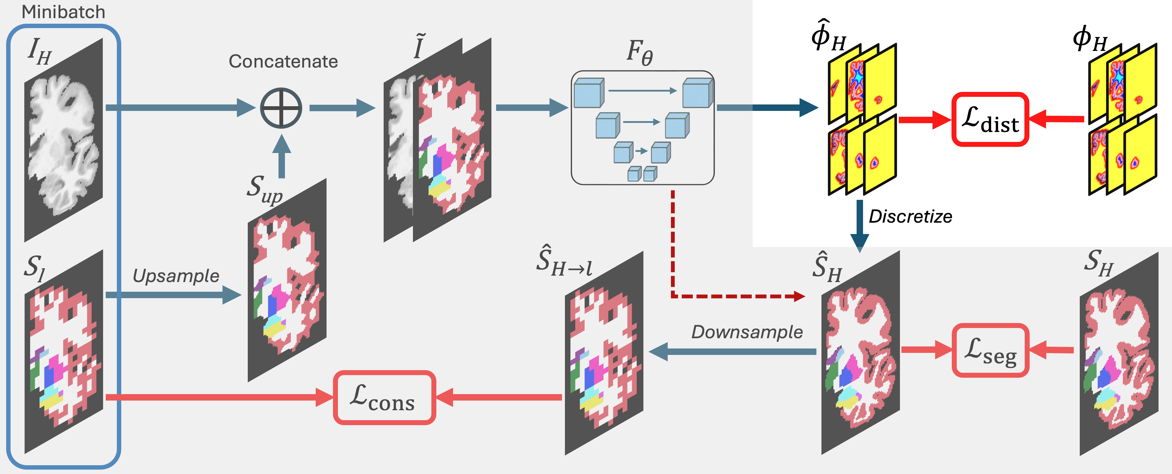
In contrast to the conventional HR-only paradigm, we propose to leverage automatically obtained LR segmentations as auxiliary supervisory signals. In human brain MRI, LR anatomical maps can be efficiently generated using robust and general-purpose segmentation tools such as SynthSeg 3, which require no manual annotation. Although coarse, these maps capture meaningful global spatial priors that can be propagated into UHR training.
Our approach integrates into training in two complementary ways, forming the first key contribution of our framework, LR-guided supervision.
Prior-Guided Input Augmentation. We first upsample the LR segmentation to the HR space via trilinear interpolation on the one-hot encoding (Figure 1, left):
and concatenate it with the raw MRI volume:
This augmented input explicitly encodes semantic context from , enabling the model to localize fine structures while retaining a global understanding of brain anatomy. Intuitively, provides a coarse anatomical atlas that conditions the network toward more plausible segmentation hypotheses. While upscaling up front is less memory efficient than in later stages, it enables compatibility with LR inputs of any size.
Cross-Resolution Semantic Consistency. In parallel, we enforce a cross-resolution alignment between predicted HR segmentations and their LR counterparts. The model output is downsampled to the coarse scale:
and compared against the reference using a Dice consistency loss:
The total loss is thus:
| (1) |
where controls the strength of cross-resolution regularization. This auxiliary loss effectively aligns the model’s HR predictions with coarse global priors, ensuring semantic coherence across scales.
Challenges and Motivation for Continuous Representations.
Patch-based training introduces inevitable alignment issues between UHR image regions and corresponding LR labels, particularly when spatial transformations are used in augmentation. Small misalignments can corrupt consistency supervision. More fundamentally, direct voxel-level classification imposes rigid, discrete boundaries, making optimization unstable and insensitive to geometric smoothness. Such formulations often yield noisy, discontinuous, or topologically inconsistent segmentations, an undesirable property when reconstructing fine anatomical interfaces.
To overcome these limitations, we reformulate the segmentation task from a categorical labeling problem into a continuous geometric regression problem, leading to our geometry-aware signed distance transform learning.
3.2 Learning Geometry-Aware Representations via Signed Distance Transforms
Definition. Given a 3D multi-class segmentation map , where each voxel is assigned a class label, the signed distance map for each class is defined as:
where, is the foreground region for class , denotes the boundary of , and is the Euclidean distance in 3 space. The full multi-class signed distance map can be represented as a tensor , where corresponds to the distance map for class .
Rather than predicting discrete voxel labels, we train the network to regress these continuous signed distance maps. This design introduces several distinct advantages over classical segmentation. First, SDFs represent spatial proximity to anatomical boundaries, capturing both interior and exterior geometry. This continuous representation yields smoother gradients and inherently encodes shape priors. Second, Distance regression provides stable optimization even in regions of partial volume or fuzzy boundaries, an essential property for submillimeter brain structures. Third, because distance fields vary smoothly across space, they naturally tolerate small misalignments or label noise, particularly when supervision comes from LR This constitutes a significant shift from discrete classification toward geometry-aware continuous learning for UHR segmentation.
Learning Formulation.
The network predicts UHR distance maps:
and is supervised with an regression loss:
We convert to probabilistic segmentations via a temperature-controlled softmax over negative distances:
Here, denotes the predicted signed distance at voxel for class , is the temperature parameter (controls the sharpness of the distance-to-probability mapping), is the probability that voxel belongs to class , and the output is a probabilistic segmentation map. This mapping smoothly bridges continuous distances and categorical probabilities, ensuring differentiability and interpretability.
Geometry-Aware Regularization.
To further enhance the geometric fidelity of learned SDFs, we introduce two additional regularizers:
The gradient norm term () enforces the unit-gradient property of true SDFs 49, while total variation (TV, ) regularization suppresses spurious local noise and promotes smooth boundary transitions. Together, they impose strong geometric priors that stabilize training and improve generalization.
Finally, we incorporate the cross-resolution consistency term from Section 3.1.2:
and define the complete loss as:
| (2) |
The architecture is illustrated in Figure 1. At inference, segmentation labels are obtained via .
This formulation bridges discrete semantic segmentation and continuous shape modeling. It encodes boundary geometry directly within the learning target, significantly improving smoothness, robustness, and topological integrity for UHR brain segmentation. In combination with LR consistency, this produces fine, anatomically coherent predictions even with limited annotations.
3.3 Scalable Class-Conditional Segmentation (SCCS)
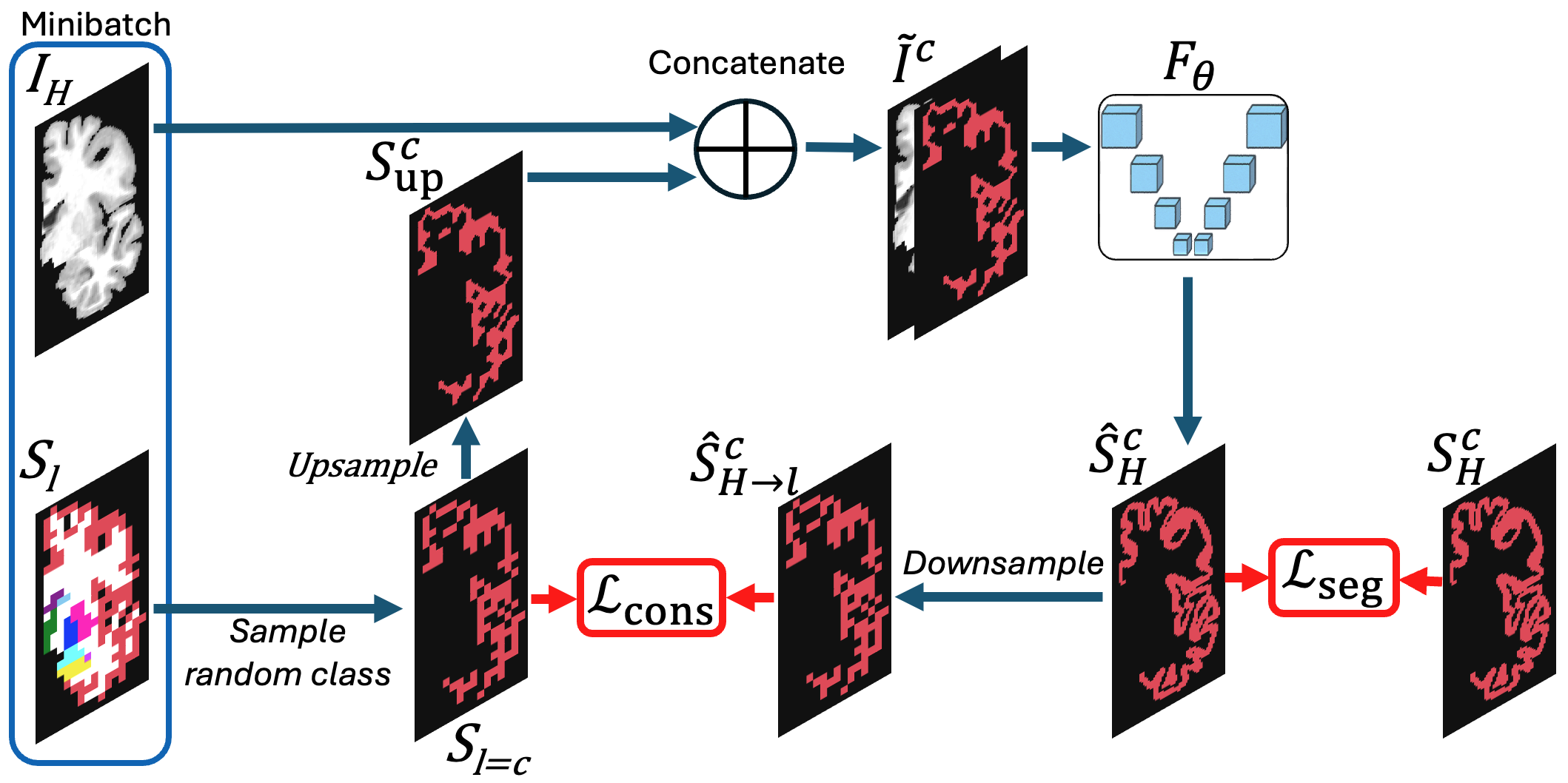
While the distance-based representation addresses geometric and boundary limitations, scaling segmentation to a large number of structures introduces additional computational bottlenecks. Standard multi-class networks must allocate one output channel per label, making them prohibitively memory-heavy for UHR volumes containing dozens or hundreds of anatomical regions.
To overcome this, we propose scalable class-conditional segmentation (SCCS), which reformulates multi-class segmentation as a collection of class-specific subproblems.
Class-Conditional Training.
Instead of predicting all classes jointly, the model learns to segment one class at a time, conditioned on a class-specific input. At each iteration, we randomly sample a target class and extract its binary mask from the LR reference :
then concatenate it with the input image:
This conditioning localizes the model’s attention to anatomically relevant regions, simplifying learning and improving sample efficiency. The model predicts a binary segmentation map and is trained via:
The total per-class loss is
| (3) |
and the whole framework is illustrated in Figure 2.
Inference and Scalability.
At inference time, the model is applied for each individual class using a class-conditioned input , which includes the image and the class-specific conditioning signal. The model produces a single-channel response map , indicating the probability that each voxel belongs to class . After looping through all classes, the individual response maps are stacked to form a multi-class prediction volume. The final multi-class segmentation is reconstructed via:
This design scales linearly with the number of classes and can seamlessly adapt to unseen anatomical labels by conditioning on their corresponding LR masks, without any retraining or architectural modification.
Generalizability via Domain Randomization. For our training paradigms, robust generalization is essential, as both the HR input and the LR guidance may originate from diverse scanners and acquisition protocols. To prevent the model from overfitting to a narrow appearance distribution, we incorporate a domain randomization strategy during training. Following recent successes in synthetic neuroimage pipelines 3; 4; 32; 37, we apply aggressive, stochastic perturbations to image contrast, noise, bias fields, and spatial resolution at each iteration.
This randomized augmentation forces the network to rely on stable structural cues rather than dataset- or scanner-specific appearance details. When combined with the class-conditional input , the model learns class-specific geometry that is inherently invariant to domain shifts. In practice, this significantly improves the model’s robustness across heterogeneous datasets and helps bridge the appearance gap between the coarse LR reference and the UHR target.
4 Experiments
We conduct comprehensive evaluations on both synthetic and real-world datasets with human-level annotations to demonstrate the effectiveness of the proposed methods as well as the effectiveness of the parameter selection.
Datasets. We use synthetic data for training, where 400 UHR isotropic scans with a resolution of mm mm mm, and the other 100 for validation. For evaluation, we employ two test sets: (1) a synthetic test set comprising 100 held-out synthetic volumes, and (2) 20 real scans from the U01 dataset 57, each with a single annotated 2D slice. The U01 surface models were originally generated by converting segmentation probability maps, obtained using a cascaded multi-resolution U-Net 69, into pseudo T1-weighted scans, followed by surface placement using a modified version of the FreeSurfer recon-all pipeline 18; 21. More details are provided in the supplementary material.
Implementation Details. We use a standard 3D U-Net 58; 17 as the backbone for our segmentation. The networks are randomly initialized and trained from scratch. We use the Dice loss 62 as segmentation loss and loss as the consistency loss (if applicable) to supervise the training of the network. Adam optimizer 43 is adopted with a learning rate of . We set the loss weights of , and as 0.1, 0.01, and 1 for all our experiments except for the ablation study sections regarding these parameters. Note that the reported memory usage corresponds to an input size of with a batch size of 1. More information is provided as supplementary material.
Baselines. CascadePSP 14 adopts a cascaded pyramid refinement strategy in which a coarse segmentation is progressively refined across multiple resolution stages. This hierarchical framework is highly effective for natural image segmentation, where dense pixel-level annotations are available and texture cues are informative. CRM 61 (Continuous Refinement Model) addresses resolution inconsistencies by explicitly fusing multi-scale feature representations to enhance boundary precision.
Evaluation Metrics. We use Dice score 73 and the Hausdorff distance (HD95) to report all the performances. The Dice score 73 is a classical segmentation metric, which measures the overlap between predicted and ground truth masks. The HD95 35 calculates the 95th percentile of all boundary distances rather than the absolute maximum. We report both means and standard deviations for all the results, and bolded numbers denote significant differences (t-test, ).
4.1 Supervised Segmentation with LR Guidance
We begin by evaluating the proposed supervised segmentation framework under various training configurations, including direct segmentation and distance-transform regression. To contextualize our approach, we compare against two state-of-the-art (SOTA) segmentation models originally developed for HR natural image segmentations: CascadePSP 14 and CRM 61. These methods represent strong HR baselines that emphasize multi-scale refinement and cross-resolution fusion, respectively, making them ideal points of comparison for assessing our design in the neuroimaging domain.
| Method | Guidance | Synthetic dataset | U01 | |||
|---|---|---|---|---|---|---|
| Dice | HD95 (mm) | Dice | HD95 (mm) | |||
| Naive seg. (Section 3.1.1) | N/A | 0.754 0.022 | 0.886 0.153 | 0.721 0.046 | 1.471 0.204 | |
| CascadePSP 14 | N/A | 0.747 0.031 | 0.901 0.161 | 0.709 0.051 | 1.515 0.228 | |
| CRM 61 | N/A | 0.761 0.019 | 0.871 0.146 | 0.719 0.049 | 1.501 0.217 | |
| Seg. (Section 3.1.2) | 0.771 0.023 | 0.803 0.124 | 0.743 0.037 | 1.015 0.198 | ||
| SDF (Section 3.2) | 0.782 0.015 | 0.768 0.106 | 0.751 0.026 | 0.976 0.151 | ||
Results. Table 1 summarizes the quantitative results across synthetic and real datasets, while qualitative examples are illustrated in Figure 3. Incorporating LR guidance consistently improves segmentation accuracy and boundary precision across all settings. Notably, the proposed SDF regression yields the highest Dice scores and lowest HD95 distances, surpassing both traditional baselines and direct segmentation models. This demonstrates that learning continuous, geometry-aware representations provides a stronger supervisory signal than discrete voxel classification, particularly when training data are limited.
 |
 |
 |
 |
 |
 |
 |
| (a) Input | (b) LR guidance | (c) GT | (d) Naive Seg | (e) Predict | (f) Regress | (g) SCCS |
Compared with CascadePSP and CRM, our approach shows superior generalization and boundary stability. Whereas the natural-image models focus on iterative refinement of visual textures, our method leverages structural priors and geometric regularization to capture the true anatomical topology of brain regions. This difference is particularly evident in high-curvature or thin cortical regions, where SDF-based learning preserves connectivity and reduces spurious discontinuities. Together, these results highlight that coupling LR anatomical guidance with geometry-aware regression offers a more robust and scalable solution for UHR brain segmentation.
4.1.1 Ablation Study
We conduct a series of ablation studies to justify the effectiveness of individual components, as well as the sensitivity to hyperparameters.
| Method | Setting | Dice | HD95 (mm) |
|---|---|---|---|
| Predict seg. | Seg. as input (w/o ) | 0.765 0.018 | 0.841 0.145 |
| Seg. as input (w/ ) | 0.771 0.023 | 0.803 0.124 | |
| Regress SDF | Seg. as input (w/o ) | 0.773 0.016 | 0.826 0.156 |
| Seg. as input (w ) | 0.782 0.015 | 0.768 0.106 |
| Dice | HD95 (mm) | |
|---|---|---|
| 0 | 0.766 0.016 | 0.812 0.124 |
| 0.05 | 0.775 0.018 | 0.826 0.110 |
| 0.10 | 0.782 0.015 | 0.768 0.106 |
| 0.15 | 0.778 0.021 | 0.756 0.129 |
| 0.20 | 0.772 0.017 | 0.801 0.098 |
| Dice | HD95 (mm) | |
|---|---|---|
| 0 | 0.768 0.019 | 0.847 0.136 |
| 0.001 | 0.765 0.022 | 0.825 0.101 |
| 0.01 | 0.782 0.015 | 0.768 0.106 |
| 0.02 | 0.779 0.010 | 0.771 0.085 |
| 0.05 | 0.766 0.021 | 0.871 0.123 |
| Dice | HD95 (mm) | |
|---|---|---|
| 0 | 0.773 0.016 | 0.826 0.156 |
| 0.5 | 0.777 0.021 | 0.746 0.090 |
| 1.0 | 0.782 0.015 | 0.768 0.106 |
| 1.5 | 0.781 0.019 | 0.790 0.141 |
| 2.0 | 0.775 0.017 | 0.782 0.161 |
Ablation Study on the Consistency Loss Term. As described earlier, the LR segmentation not only provides spatial guidance but also enforces semantic alignment between the predicted HR output and its LR reference. To assess the impact of the cross-resolution consistency term , we perform an ablation study by removing it from the overall objective. The results in Table 2 show a clear and consistent drop in Dice score and an increase in boundary error when is omitted. This confirms that the consistency constraint effectively regularizes the network, promoting spatial coherence and improving generalization across varying resolutions.
| Dice | HD95 (mm) | |||
|---|---|---|---|---|
| ✗ | ✗ | ✗ | 0.748 0.025 | 1.023 0.176 |
| ✓ | ✗ | ✗ | 0.765 0.018 | 0.937 0.181 |
| ✗ | ✓ | ✗ | 0.760 0.012 | 0.895 0.161 |
| ✗ | ✗ | ✓ | 0.754 0.021 | 1.123 0.145 |
| ✓ | ✓ | ✗ | 0.773 0.016 | 0.826 0.156 |
| ✓ | ✗ | ✓ | 0.768 0.019 | 0.847 0.136 |
| ✗ | ✓ | ✓ | 0.766 0.016 | 0.812 0.124 |
| ✓ | ✓ | ✓ | 0.782 0.015 | 0.768 0.106 |
Ablation Study on Loss Weights. We next examine the sensitivity of the proposed framework to the weighting coefficients in the total loss formulation (Equation 2). Specifically, we vary the relative strengths of , , and as reported in Table 5, Table 5, and Table 5. The results indicate that performance remains stable across a wide range of values, suggesting that the framework is not overly sensitive to hyperparameter tuning. This robustness simplifies model training and supports the practical applicability of our approach across datasets with different contrast and noise characteristics.
Ablation Study on the Loss Components. Finally, we evaluate the individual and joint contributions of each loss component in Equation 2. As summarized in Table 6, removing any single term leads to a measurable degradation in performance, whereas combining all components yields the highest Dice accuracy and the lowest boundary error. This confirms that the gradient norm (), total variation (), and consistency () terms are complementary: the first two enforce geometric regularity and smoothness, while the latter maintains cross-resolution alignment. Together, they guide the network toward anatomically coherent, high-fidelity segmentations.
4.2 Scalable Class-Conditional Segmentation (SCCS)
We further evaluate the effectiveness and scalability of the proposed SCCS strategy through controlled experiments.
Comparison with Classical Multi-class Segmentation. Table 7 presents a comparison between the proposed SCCS framework and the conventional multi-class segmentation setting, where all class-conditioning channels are processed jointly. The classical all-class model achieves a comparable Dice score (0.771 0.023 vs. 0.769 0.016) but requires higher GPU memory (particularly for a high number of classes) and cannot readily generalize to new, previously unseen classes. In contrast, SCCS focuses on one class at a time, resulting in a constant memory footprint that does not scale with the number of classes. This property makes SCCS particularly well-suited for UHR segmentation tasks involving numerous anatomical structures or when hardware resources are limited.
| Method | Input Channels | Dice | HD95 |
|---|---|---|---|
| All-class conditioning | 0.771 0.023 | 0.803 0.124 | |
| SCCS (Ours) | 0.769 0.016 | 0.798 0.117 |
Generalization to Held-out Classes. To further assess flexibility and generalization, we evaluate SCCS on unseen anatomical classes. Specifically, one class is excluded during training and later introduced only at test time, as summarized in Table 8. While the conventional multi-class model performs slightly better on seen classes (0.782 0.019 vs. 0.771 0.016), it is fundamentally restricted to the fixed set of labels used during training and cannot infer new structures without retraining. In contrast, SCCS, by design, accepts a class-conditional input that specifies the target class, enabling it to segment previously unseen structures directly. Despite not having encountered the held-out class during training, SCCS achieves a reasonable Dice score of 0.687 0.036, demonstrating its capacity to generalize across classes. This property is particularly valuable in evolving neuroimaging datasets where anatomical definitions, label sets, or study protocols may expand over time. By decoupling segmentation from fixed label dependencies, SCCS provides a flexible, scalable, and future-proof solution for UHR anatomical segmentation.
| Method | Input Channels | Seen Classes (Dice ) | Unseen Classes (Dice) |
|---|---|---|---|
| Multi-class seg. | 0.782 0.019 | N/A | |
| SCCS (Ours) | 0.771 0.016 | 0.687 0.036 |
5 Conclusion
We introduced a learning-based framework for upscaling 3D segmentations, framing resolution transfer as a general representation learning problem rather than a domain-specific anatomical task. Our method learns to infer HR semantic detail from coarse volumetric labels by predicting continuous signed distance representations, enabling accurate and geometry-aware label refinement without direct supervision at UHRs. Through class-conditional conditioning and scalable architectural design, the approach generalizes across structures and datasets while maintaining computational efficiency. Experiments on HR ex vivo MRI demonstrate that the proposed framework bridges the gap between coarse and fine segmentation regimes, offering a scalable path toward high-fidelity 3D label synthesis. More broadly, we see this as a step toward learning-based resolution transfer in structured visual data — extending the idea of label “super-resolution” beyond images to the space of semantic 3D geometry.
References
- Deep learning for brain MRI segmentation: state of the art and future directions. Journal of digital imaging. Cited by: §1.
- Distance transform regression for spatially-aware deep semantic segmentation. CVIU. Cited by: §2.
- SynthSeg: segmentation of brain mri scans of any contrast and resolution without retraining. MedIA. Cited by: §2, §3.1.2, §3.3, §7.
- Robust machine learning segmentation for large-scale analysis of heterogeneous clinical brain mri datasets. PNAS. Cited by: §2, §3.3.
- FlowSDF: flow matching for medical image segmentation using distance transforms. IJCV. Cited by: §2.
- Universeg: universal medical image segmentation. In ICCV, Cited by: §1, §2.
- Modeling the background for incremental learning in semantic segmentation. In CVPR, Cited by: §2.
- Unsupervised inference of signed distance functions from single sparse point clouds without learning priors. In CVPR, Cited by: §2.
- Transunet: transformers make strong encoders for medical image segmentation. arXiv preprint arXiv:2102.04306. Cited by: §1, §2.
- Semantic image segmentation with deep convolutional nets and fully connected crfs. arXiv preprint arXiv:1412.7062. Cited by: §2.
- Deeplab: semantic image segmentation with deep convolutional nets, atrous convolution, and fully connected crfs. TPAMI. Cited by: §2.
- Rethinking atrous convolution for semantic image segmentation. arXiv preprint arXiv:1706.05587. Cited by: §2.
- CPM: class-conditional prompting machine for audio-visual segmentation. In ECCV, Cited by: §2.
- Cascadepsp: toward class-agnostic and very high-resolution segmentation via global and local refinement. In CVPR, Cited by: §4.1, Table 1, §4.
- Neural unsigned distance fields for implicit function learning. In NeurIPS, Cited by: §2.
- Gensdf: two-stage learning of generalizable signed distance functions. In NeurIPS, Cited by: §2.
- 3D U-Net: learning dense volumetric segmentation from sparse annotation. In MICCAI, Cited by: §4.
- Cortical surface-based analysis: I. segmentation and surface reconstruction. Neuroimage. Cited by: §4.
- An image is worth 16x16 words: transformers for image recognition at scale. arXiv preprint arXiv:2010.11929. Cited by: §2.
- Interactive few-shot learning: limited supervision, better medical image segmentation. TMI. Cited by: §1, §2.
- Cortical surface-based analysis: II: inflation, flattening, and a surface-based coordinate system. Neuroimage. Cited by: §4.
- Automated segmentation and shape characterization of volumetric data. NeuroImage. Cited by: §1.
- Incremental few-shot instance segmentation. In CVPR, Cited by: §2.
- Multi-domain incremental learning for semantic segmentation. In WACV, Cited by: §2.
- Transfer learning for domain adaptation in MRI: application in brain lesion segmentation. In MICCAI, Cited by: §1.
- An image-processing system for qualitative and quantitative volumetric analysis of brain images. JCAT. Cited by: §1.
- Synthetic data in generalizable, learning-based neuroimaging. Imaging Neuroscience. Cited by: §2.
- Registration by regression (rbr): a framework for interpretable and flexible atlas registration. In WBIR, Cited by: §2.
- Deep learning enables automatic detection and segmentation of brain metastases on multisequence MRI. JMRI. Cited by: §1.
- Brain tumor segmentation with deep neural networks. MedIA. Cited by: §2.
- Fastsurfer-a fast and accurate deep learning based neuroimaging pipeline. NeuroImage. Cited by: §1, §2.
- SynthMorph: learning contrast-invariant registration without acquired images. TMI. Cited by: §2, §3.3.
- Hierarchical uncertainty estimation for learning-based registration in neuroimaging. In ICLR, Cited by: §2.
- Learn2Synth: learning optimal data synthesis using hypergradients for brain image segmentation. In ICCV, Cited by: §2.
- Comparing images using the Hausdorff distance. TPAMI. Cited by: §4.
- Corpus callosum morphology in attention deficit-hyperactivity disorder: morphometric analysis of MRI. Journal of Learning Disabilities. Cited by: §1.
- SynthSR: a public ai tool to turn heterogeneous clinical brain scans into high-resolution t1-weighted images for 3d morphometry. Science advances. Cited by: §2, §3.3.
- Multi-atlas segmentation of biomedical images: a survey. MedIA. Cited by: §1, §2.
- NnU-net: a self-configuring method for deep learning-based biomedical image segmentation. Nature methods. Cited by: §2.
- Sdfdiff: differentiable rendering of signed distance fields for 3D shape optimization. In CVPR, Cited by: §2.
- Efficient multi-scale 3D CNN with fully connected crf for accurate brain lesion segmentation. MedIA. Cited by: §1, §2.
- Automated deep learning segmentation of high-resolution 7 tesla postmortem MRI for quantitative analysis of structure-pathology correlations in neurodegenerative diseases. Imaging Neuroscience. Cited by: §1, §3.1.1, §3.1.1.
- Adam: a method for stochastic optimization. arXiv preprint arXiv:1412.6980. Cited by: §4.
- Panoptic feature pyramid networks. In CVPR, Cited by: §2.
- Segment anything. In ICCV, Cited by: §1, §2.
- Brain-id: learning contrast-agnostic anatomical representations for brain imaging. In ECCV, Cited by: §2.
- An improved implementation of brain tumor detection using segmentation based on soft computing. Journal of Cancer Research and Experimental Oncology. Cited by: §1.
- Fully convolutional networks for semantic segmentation. In CVPR, Cited by: §2.
- Towards better gradient consistency for neural signed distance functions via level set alignment. In CVPR, Cited by: §3.2.
- How distance transform maps boost segmentation cnns: an empirical study. In MIDL, Cited by: §2.
- Brain tumor target volume determination for radiation treatment planning through automated MRI segmentation. International Journal of Radiation Oncology, Biology, Physics. Cited by: §1.
- V-net: fully convolutional neural networks for volumetric medical image segmentation. In 3DV, Cited by: §1, §2.
- Learning deconvolution network for semantic segmentation. In ICCV, Cited by: §2.
- Constructing signed distance functions. Level set methods and dynamic implicit surfaces. Cited by: §2.
- Self-supervised learning for few-shot medical image segmentation. TMI. Cited by: §2.
- Deepsdf: learning continuous signed distance functions for shape representation. In CVPR, Cited by: §2.
- 3D molecular phenotyping of cleared human brain tissues with light-sheet fluorescence microscopy. Communications Biology. Cited by: §4, §7.
- U-net: convolutional networks for biomedical image segmentation. In MICCAI, Cited by: §1, §2, §4.
- QuickNAT: a fully convolutional network for quick and accurate segmentation of neuroanatomy. NeuroImage. Cited by: §2.
- Fuzzy distance transform: theory, algorithms, and applications. CVIU. Cited by: §2.
- High quality segmentation for ultra high-resolution images. In CVPR, Cited by: §4.1, Table 1, §4.
- Generalised dice overlap as a deep learning loss function for highly unbalanced segmentations. In DLMIA, Cited by: §4.
- Inferring the class conditional response map for weakly supervised semantic segmentation. In WACV, Cited by: §2.
- Recurrent mask refinement for few-shot medical image segmentation. In ICCV, Cited by: §2.
- Domain randomization for transferring deep neural networks from simulation to the real world. In IROS, Cited by: §2.
- Monitoring brain tumor response to therapy using MRI segmentation. Magnetic resonance imaging. Cited by: §1.
- Multi-modal learning from unpaired images: application to multi-organ segmentation in CT and MRI. In WACV, Cited by: §2.
- Deep distance transform for tubular structure segmentation in CT scans. In CVPR, Cited by: §2.
- Segmentation of supragranular and infragranular layers in ultra-high-resolution 7T ex vivo MRI of the human cerebral cortex. Cerebral Cortex. Cited by: §3.1.1, §4.
- Data augmentation in class-conditional diffusion model for semi-supervised medical image segmentation. In IJCNN, Cited by: §2.
- Nnformer: interleaved transformer for volumetric segmentation. arXiv preprint arXiv:2109.03201. Cited by: §2.
- Models genesis: generic autodidactic models for 3D medical image analysis. In MICCAI, Cited by: §2.
- Statistical validation of image segmentation quality based on a spatial overlap index1: scientific reports. Academic radiology. Cited by: §4.
Supplementary Material
6 Overview
In the supplementary material, we begin with the details of the datasets Section 7, followed by the implementation details in Section 8. Then, we provide the computational resources in Section 9, followed by a few other qualitative samples in Section 10. The limitations are provided in Section 11, followed by an analysis on the broader impact in Section 12 and a statement on the use of LLMs in Section 13.
7 Dataset Details
Synthetic data. As described in the main text, we use synthetic data for training. The UHR isotropic scans are generated from created segmentation labels at a resolution of mm mm mm. The coarse, LR segmentation is obtained using SynthSeg 3, which segments approximately 30 brain regions at 1 mm isotropic resolution, regardless of the input resolution. We group these regions into 7 foreground classes, Cortex, White Matter, Thalamic Mask, Pallidum Mask, Putamen Mask, Caudate and Accumbens, and Cerebellar Gray Matter, along with a background class. The detailed mapping list will be provided to ensure reproducibility.
Real data. Following the same class grouping used for the synthetic data, we asked expert annotators to manually label one representative slice from each of 20 real scans in the U01 dataset 57.
8 Implementation Details
Network details. The 3D UNet architecture employed in this paper follows an encoder-decoder structure with skip connections, designed to capture both global context and fine-grained spatial details in volumetric medical images. The encoder consists of four downsampling blocks, each composed of two 3D convolutional layers followed by batch normalization and LeakyReLU activations, with 3D max pooling used to progressively reduce spatial resolution while increasing the number of feature channels. The bottleneck (or bridge) layer connects the encoder and decoder, maintaining the deepest representation with the highest channel dimension. The decoder mirrors the encoder with four upsampling blocks, where each block begins with a transposed 3D convolution to upsample the feature map, followed by concatenation with the corresponding encoder features (skip connection), and two additional convolutional layers with normalization and activation. The final output layer is a 3D convolution that maps the feature maps to the desired number of output channels. This design enables precise voxel-wise predictions while maintaining spatial consistency across the 3D volume. The codes will be released upon acceptance to ensure reproducibility.
9 Computational Resources
The experiments are conducted on an NVIDIA A40 GPU (48GB), using a 26-core Intel(R) Xeon(R) Gold 6230R CPU @ 2.10GHz and 200 GB RAM.
10 Qualitative Results
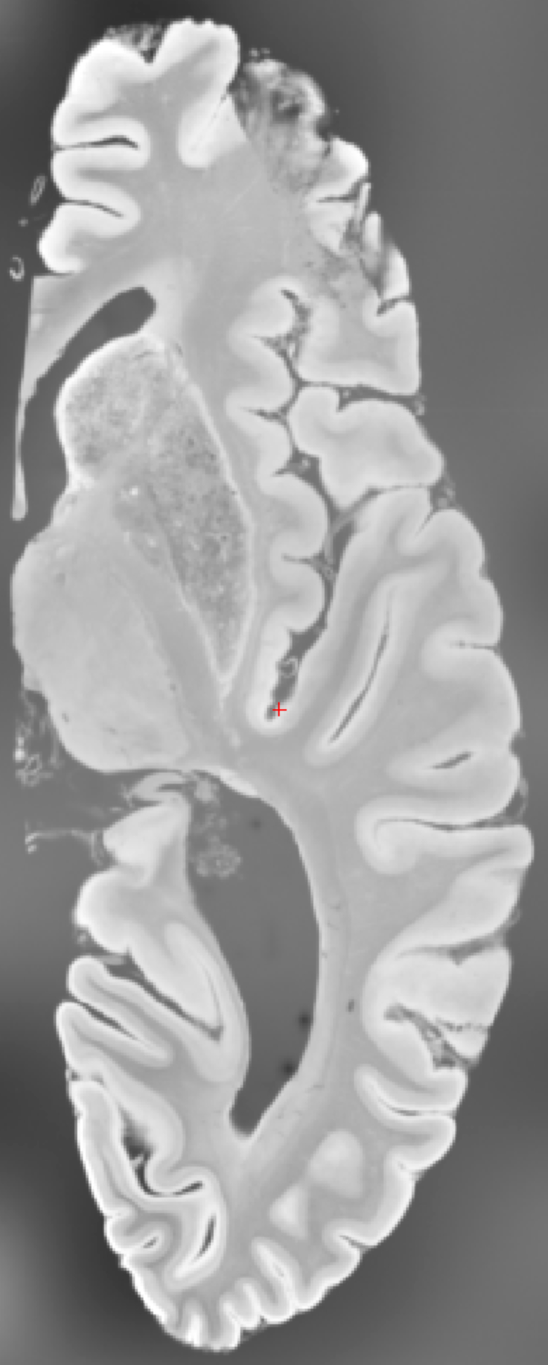
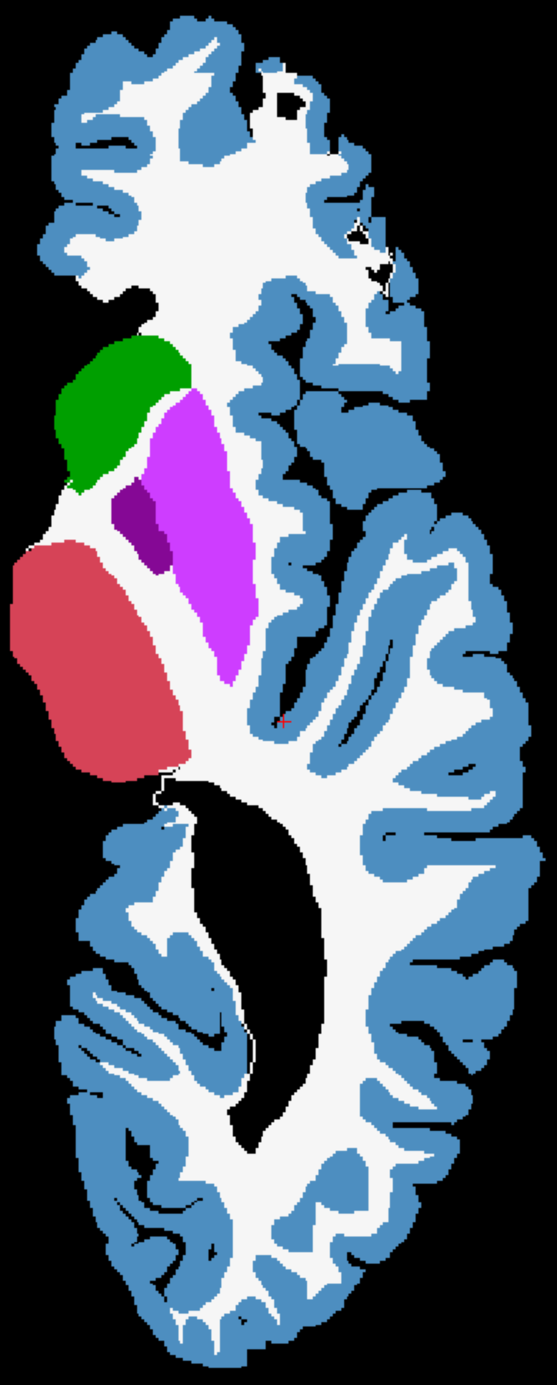
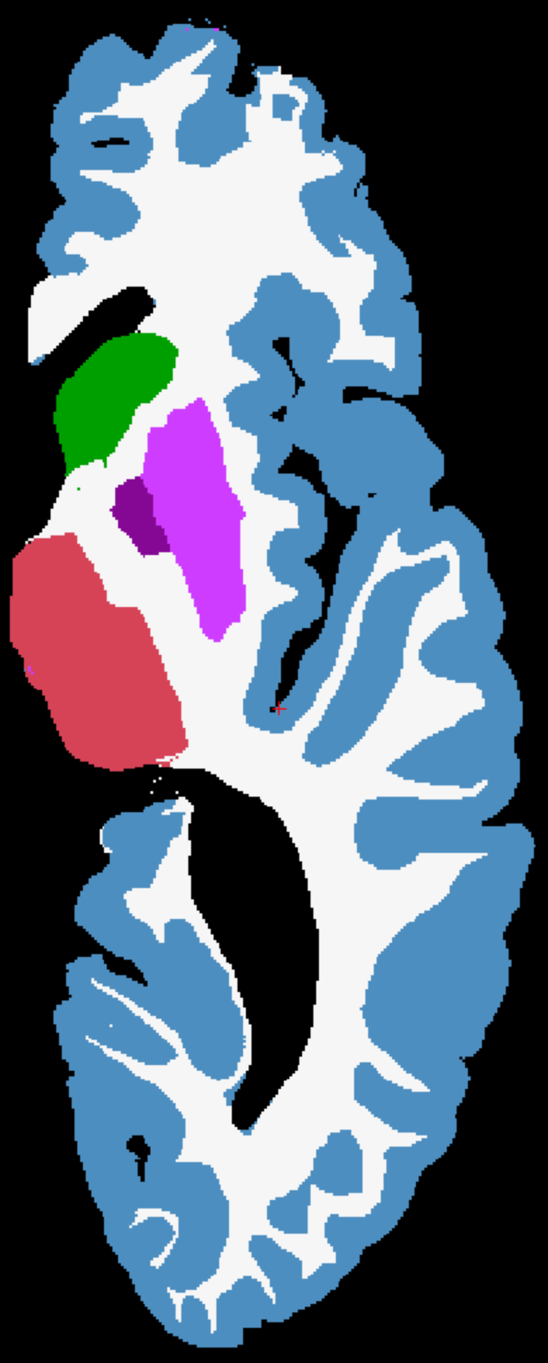
For qualitative results, we provide another sample from U01 in Figure 4.
 |
 |
 |
 |
 |
 |
 |
| (a) Input | (b) LR guidance | (c) GT | (d) Naive Seg | (e) Predict | (f) Regress | (g) SCCS |
11 Limitations
A key limitation of the proposed method lies in its reliance on automatic LR segmentations as spatial guidance. While this strategy significantly reduces manual labeling effort, it inherently assumes that these coarse labels are accurate and spatially consistent. In practice, however, these LR segmentations may contain systematic biases or anatomical imprecision. These imperfections can propagate through the network, potentially leading to degraded segmentation performance at higher resolutions. One possible solution is to incorporate uncertainty modeling or confidence-weighted supervision, where the model learns to discount or correct for less reliable regions in the coarse labels. Additionally, leveraging self-supervised refinement mechanisms that iteratively improve the alignment between LR and HR outputs could further mitigate this issue.
Another important limitation is the limited validation on real, fully annotated 3D clinical datasets. Although the paper demonstrates strong performance on synthetic data and sparsely labeled real data (e.g., single-slice annotations), the generalizability of the proposed approach to densely annotated, HR clinical scans remains uncertain. This is particularly relevant given the variability in acquisition protocols, scanner hardware, and anatomical differences across patient populations. To strengthen the empirical evidence and assess robustness, future work should include comprehensive benchmarking on public and private datasets with full volumetric annotations (e.g., HCP, OASIS, ADNI). Incorporating domain adaptation techniques or semi-supervised learning frameworks could also help bridge the gap between synthetic and clinical domains, further enhancing the method’s practical utility in real-world settings.
12 Broader Impact
The broader impact of our work lies in its potential to democratize access to detailed, high-fidelity brain image analysis without the prohibitive cost of dense manual annotations. By leveraging LR coarse labels and a scalable, class-conditional framework, the proposed method makes it feasible to segment UHR brain MR scans, which are increasingly used in neuroscience and clinical research, using limited supervision and computational resources. This can accelerate research in neurodegenerative diseases, brain development, and population-level studies where large-scale, accurate segmentation is essential.
Additionally, the framework’s ability to generalize to unseen classes and operate efficiently in memory-constrained settings makes it adaptable to LR clinical environments or global health applications. However, as with any medical AI tool, careful validation is essential to avoid biases or errors introduced by synthetic or weak labels. If responsibly developed and adopted, the method could contribute meaningfully to advancing scalable, accessible, and precise neuroimaging analysis.
13 Usage of LLM
We only use LLM to improve the writing quality and grammar check of the manuscript.